Hand Surgery Source

BRACHIAL PLEXUS INJURY

Introduction

Brachial plexus injuries are considered one of the most severe and devastating nerve injuries affecting the upper extremity. Most brachial plexus injuries are closed traumatic injuries resulting from motorcycle accidents that damage the upper extremity nerves by forcing the shoulder and head apart. These traumatic brachial plexus injuries are the most common type of brachial plexus injury.  However, open brachial plexus injuries and neonatal brachial plexus injuries are also possible. Patients that sustain these injuries typically experience loss of sensation, loss of motor power in some or all of the affected upper extremity and develop significant neuropathic pain. These symptoms can complicate or completely preclude many motor tasks that involve the impaired shoulder, elbow, wrist or hand. Conservative treatment consisting of careful observation may be adequate for some brachial plexus injuries in which the damage is not severe, for example after low velocity gunshot wounds. However surgery is often needed for more severe injuries like nerve avulsion injuries that cut or rupture all or some neural components of the plexus. Surgery may be initiated immediately or delayed under certain circumstances.  Treatment options include direct nerve repair, nerve grafting, nerve transfers, tendon transfer, muscle transfer, joint arthrodesis and neurolysis.1,2,13,15

Pathophysiology

  • Closed brachial plexus injuries are usually associated with a traction mechanism where the arm and shoulder are forcefully distracted away from the neck or trunk.  This mechanism of injury commonly results in root avulsion lesions.13,15
    • The majority of these injuries occur in motorcycle or snow mobile accidents. When the individual falls to the ground and the head and shoulder are forced apart, damage is caused to the brachial plexus.2,3
    • Other less common mechanisms include nerve crush or compression injuries caused by car accidents and in contact sports2
    • Open brachial plexus injuries typically result from stab wounds, gunshot wounds, other instruments and missiles, and occasionally open fractures of the shoulder girdle
      • These types of traumatic brachial plexus injuries are far less common than closed brachial plexus injuries2
      • Lower brachial plexus injuries are characterized by damage to the C7, C8, or T1 nerve roots, which results in dysfunction of the intrinsic muscles of the hand, wrist flexors, and the flexors and extensors of the fingers and thumb4
      • Neonatal brachial plexus injuries are also possible, and shoulder dystocia is the most common mechanism of injury in these cases. Sixty percent of these neonatal brachial plexus injuries are mild and spontaneously resolve, while more severe injuries often have longer-term dysfunction with varying degrees of restricted function involving the shoulder, arm, or hand5 (Also see - the diagnostic guide for brachial plexus birth injury.)

Related Anatomy

  • Spinal cord with motor neurons15
  • Ventral rami
  • Dorsal rami with dorsal root ganglion containing sensory neurons
  • Cervical Roots
    • The brachial plexus is formed by the cervical roots C5, C6, C7, C8 and T1. Each root is formed by ventral and dorsal rami that leave the cervical spinal cord via the cervical spinal foramen. The roots then become the trunks of the brachial plexus. The trunks divide into the anterior and posterior divisions which divide and combine to form the lateral, posterior and medial cords. Branching off the roots, upper trunk and cords are the nerves of the upper extremity13-15 A brachial plexus originating from C5-T1 is the common anatomical makeup. If C4 contribute, it is called a prefixed plexus and if T2 contribute to the plexus, it is called a postfixed plexus15
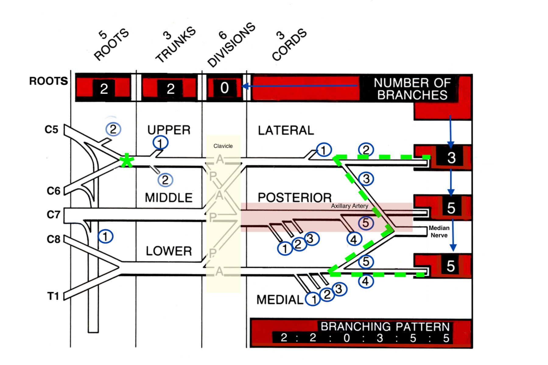
  • The brancing of the nerves of the bradchial plexus follows the Nerve Branching Rule 2-2-0-3-5-5 (modified Last's Rule).
    • (2) Two nerve branches arise from the roots of the brachial plexus:
      • Long thoracic nerve which innervates the serratus anterior
      • Dorsal scapular nerve which innervates the rhomboid and levator scapulae
      • Note: The phrenic nerve arises from the cervical plexus (C6, C4 and C5). The majority of the phrenic nerve come from C4 but some axons originate from the C5 root which is a component of the brachial plexus.
    • (2)  Two nerve branches arise from the upper trunk of the brachial plexus: 
      • Subclavius nerve which innervates the subclavius muscle
      • Suprascapular nerve which innervateds gthe supraspinatus and infraspinatus muscles.
    • (0)  No nerve branches arise from the anterior or posterior divisions of the brachial plexus. These divisions lie posterior to the clavicle. The anterior       divisions ultimately supply the flexor muscles of the upper extremity and the posterior divisions supply the extensor muscles..
    • (3)  Three nerve branches originate from the lateral cord of the brachial plexus:
      • Musculocutaneous nerve which innervates the biceps, brachialis and coracobrachialis.
      • Lateral head of the median nerve
      • Lateral pectoral nerve which innervates the pectoralis major muscle
    • (5)  Five never branches originate from the posterior cord of the brachial plexus:
      • Upper subscapular nerve which innervates the subscapularis muscle
      • Thoracodorsal nerve which innervates the lattisimus dorsi muscle
      • Lower subscapular nerve which innervates the subscapularis and teres major muscles
      • Axillary nerve which innervates the deltoid and teres minor muscles
      • Radial nerve which innervates the triceps, brachioradialis, wrist, finger and thumb extensor
    • (5)  Five nerve branches originate from the medial cord of the brachial plexus:
      • Medial pectoral nerve which innervates the pectoralis major and minor muscles
      • Medial cutanous nerve of the arm which provides sensation to the medial arm
      • Medial cutaneous nerve of the forearm which provides sensation to the medial forearm
      • Ulnar nerve which innervates the FCU, FDP IV and V and ulnar intrinsic muscles of the hand
      • Medial head of the median nerve
  • Sympathetic ganglion
  • Erb’s point - the point where the C5 and C6 roots combine to form the upper trunck 2-3cm above the clavicle.  Injuries at Erb's point damage the axillary, musculocutaneous, and suprascapular nerves.
  • Brachial plexius "M" shape - When dissecting the brachial plexus, a useful landmark is the nerves' "M" pattern on the supeficial surface of the axillary artery.  This "M" shape is formed by the nusculocaneous nerve, median nerve, and ulnar nerve.  These nerves lie posterior to the pectorais muscle and just distal to the clavicle and the brachial plexus anterior and posterior divisions.
  • Scalenus anterior muscle - as the nerves of the brachial plexus enter the upper extremity they pass between the scalene muscles.
  • Scalenus medius muscle
  • Scalenus posterior muscle
  • Pectorais major muscle
  • Pectorais minor muscle

Brachial Plexus Injury Classicications

  • Brachial plexus injuries are commonly grouped using the Seddon's classification systems of peripheral nerve injuries into neurapraxia, axonotmesis, and neurotmesis, along with Sunderland’s first-to-fifth-degree injury categories
    • Neurapraxia (first-degree injury)
      • Localized myelin damage and conduction deficiencies
      • Complete spontaneous recovery could be expected in 4-12 weeks
  • Axonotmesis (second-degree injury)
    • Disruption of the nerve cell’s axon, followed by Wallerian degeneration
    • Complete axonal regeneration could be expected to occur at a rate of approximately 1-3 mm/day from the injury site to the target muscle
  • Third-degree injury
    • Internal derangement of the endoneurium and intrafascicular fibrosis precludes complete regeneration and results in partial recovery
  • Fourth-degree injury
    • Due to perineurial and fascicular disruption, neuroma-in-continuity forms a complete scar block and spontaneous recovery is not expected
  • Neurotmesis (fifth-degree injury)
    • Complete nerve transection with the need for surgical intervention6,7

Another classification system divides brachial plexus injuries into two groups according to the site of injury's relationship to the clavicle

  •  Supraclavicular lesions
    • Implies injury at the spinal nerve and trunks levels
  • Infraclavicular lesions
    • Typically occur at the cord and terminal branch levels2
    • Many experts also find that further subdividing brachial plexus injuries into preganglionic and postganglionic lesions can be beneficial when making treatment palns for patients with brachual plexus injuries.
      • Preganglionic lesions
        • Occur proximal to the dorsal root ganglion and involve the central nervous system (CNS)
        • Have a poor prognosis due to the inability of CNS nerves to regenerate
        • Sensation is absent, but sensory nerve action potentials (SNAP's) will be intact on EMG because injury is proximal to the dorsal nerve cell bodies in the dorsal rami.
      • Postganglionic lesions
        • Occur distal to dorsal root ganglion and involve the peripheral nervous system (PNS )
        • Have a better prognosis owing to PNS nerve regenerative ability
        • Sensation and SNAP's absent on EMG testing15

Incidence and Related Conditions

  • The exact number of annual brachial plexus injuries is difficult to estimate, but their incidence has been rising in recent years due to increases in extreme sport participation and motor vehicle accident survivors.  There is a significant predilection in male gender and individuals aged 15-25 years old8
  • ~70% of traumatic brachial plexus lesions are due to traffic accidents and 70-80% of these result from motorcycle accidents9 and snow mobile accidents13-15
  • 70-75% of traumatic brachial plexus injuries are located in the supraclavicular region, with 75% of these involving total plexus lesions (C5-T1), 20-25% involving C5-C6 root injuries, and 2-3.5% involving isolated C8-T1 root lesions10
  • Total brachial plexus injuries usually involve rupture of C5-C6 roots and avulsion of C7-T1 roots10
  • The overall incidence and types of brachial plexus injuries is well summarized by Naraka's Law of Seven 70's 9,15
    1. 70% secondary MVA
    2. 70% of MVA secondary to motorcycles
    3. 70% have the patients with brachial plexus injuries have other injuries
    4. 70% have a supraclavicular component to their injury
    5. 70% have at least one root avulsion
    6. 70% involved C7 if root avulsed
    7. 70% have persisted neuropathic pain

Differential Diagnosis

  • Acromioclavicular joint injury
  • Cervical disc injury
  • Cervical discogenic pain syndrome
  • Cervical radiculopathy
  • Cervical sprain/strain
  • Partial Brachial Plexus Injuries
    • Erb palsy - this is an injury to the upper trunk of the brachial plexus. It damages the axillary, musculocutaneous, and suprascapular nerves.
    • Klumpke palsy - this is an injury to the lower roots of the brachial plexus. It damages the muscle function in the forearm, wrist and hand. This injury is associated with the waiter's tip sign because the elbow is extended, forearm pronated, wrist flexed and fingers partially flexed. 
  • Parsonage Turner syndrome
  • Shoulder dislocation
  • Shoulder impingement syndrome
  • Thoracic outlet syndrome
ICD-10 Codes

BRACHIAL PLEXUS INJURY

Diagnostic Guide Name

BRACHIAL PLEXUS INJURY

ICD 10 Diagnosis, Single Code, Left Code, Right Code and Bilateral Code

DIAGNOSIS SINGLE CODE ONLY LEFT RIGHT BILATERAL (If Available)
BRACHIAL PLEXUS INJURY S14.3XX_      

Instructions (ICD 10 CM 2020, U.S. Version)

THE APPROPRIATE SEVENTH CHARACTER IS TO BE ADDED TO EACH CODE FROM CATEGORY S14
A - Initial Encounter
D - Subsequent Routine Healing
S - Sequela

ICD-10 Reference

Reproduced from the International statistical classification of diseases and related health problems, 10th revision, Fifth edition, 2016. Geneva, World Health Organization, 2016 https://apps.who.int/iris/handle/10665/246208

Basic Science Photos and Related Diagrams
Brachial Plexus Related Diagrams
  • Brachial Plexus diagram showing roots, trunks, divisions & cords. (Remember RTDCBs - Running Together Down Country Byways). Also note the Nerve Branching  2 2 0 3 5 5 Rule for the number of branches from each part of the plexus. Also see the brachial plexus branching table in the next image. Note the green asterisk at Erb’s point.  Also note the brachial plexus “M” landmark shape anterior to the axillary artery which is defined by the musculocutaneous nerve, the median nerve and the ulnar nerve. Finally not
    Brachial Plexus diagram showing roots, trunks, divisions & cords. (Remember RTDCBs - Running Together Down Country Byways). Also note the Nerve Branching 2 2 0 3 5 5 Rule for the number of branches from each part of the plexus. Also see the brachial plexus branching table in the next image. Note the green asterisk at Erb’s point. Also note the brachial plexus “M” landmark shape anterior to the axillary artery which is defined by the musculocutaneous nerve, the median nerve and the ulnar nerve. Finally note the divisions of the brachial plexus posterior to the clavicle.
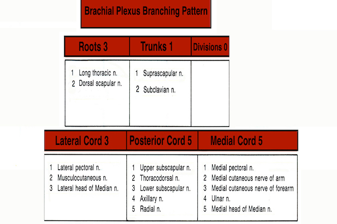
  • Brachial Plexus Branching Pattern
    Brachial Plexus Branching Pattern
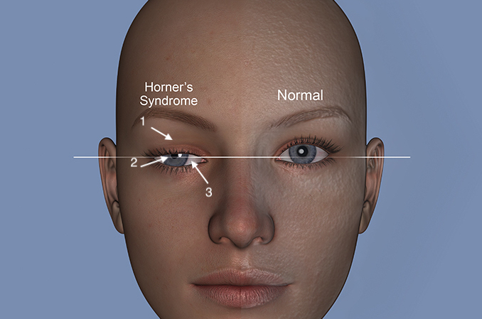
  • Horner’s sign is frequently associated with C8 and T1 root avulsions which also damage the sympathetic fibers to the face and may produce an ipsilateral Horner’s sign: 1) Ptosis (drooped eyelid); 2) meiosis (constricted pupil); 3) enophthalmos (sunken globe).  Horner’s sign is associated with anhydrous of the ipsilateral face and a poor prognosis when connected with a brachial plexus injury.
    Horner’s sign is frequently associated with C8 and T1 root avulsions which also damage the sympathetic fibers to the face and may produce an ipsilateral Horner’s sign: 1) Ptosis (drooped eyelid); 2) meiosis (constricted pupil); 3) enophthalmos (sunken globe). Horner’s sign is associated with anhydrous of the ipsilateral face and a poor prognosis when connected with a brachial plexus injury.
Symptoms
History of brachial plexus injury e.g. motorcycle accident
Persistent upper extremity pain
Flail arm and/or stiffness in one or more upper extremity joints
Loss of elbow flexion and/or extension, loss of wrist extension and/or loss of shoulder control
Typical History

The typical patient is a 28-year-old male who sustained a brachial plexus injury during a motorcycle accident. During the accident, the man fell to the ground with his arms outstretched, which caused a forced separated his head and right shoulder.  The impact dislocated the shoulder and causing severe stretching injury to the right brachial plexus. He was unable to move his right shoulder, elbow, or hand, and reported numbness and a burning sensation in several locations. This substantially impaired his ability to perform any tasks requiring his injured arm. He also ruptured his spleen, fractured three ribs and fractured his left tibia. He was treated at the scene by EMS and brought to a level 1 trauma center by helicopter. Three months after his accident, he underwent reconstructive surgery for his right brachial plexus.

Positive Tests, Exams or Signs
Work-up Options
Treatment Options
Treatment Goals
  • Restore upper extremity function by first obtaining elbow flexion and second by obtaining active shoulder stabilization. Obtaining hand and wrist function remains an elusive third goal after brachial plexus injury.
  • Minimize neuropathic pain

Three goals that are specific to patients with brachial plexus injuries are13,15:

  1. Identifying the patients who will benefit from surgery
  2. Timing the surgery remains controversial. Operating too early may mean doing procedures on patients who will recover spontaneously. Operating too late may not be successful because the muscles may me degenerated and unable to benefit from reinnervation.
  3. Prioritizing the functions to be restored. As noted, most surgeons try to get elbow flexion first, shoulder stabilization second and forearm/hand function third.
Conservative
  • Conservative management consisting of close, regular observation is recommended when damage from the brachial plexus injury is not complete. Obersavation is also appropriate for low velocity gunshot wounds that do not cause any major vascular damage. These injuries may recover spontaneously1,3  A conservative approach is also indicated if the palsy was incurred from carrying a heavy rucksack, by malpositioning during an operation, or during sleeping.1
  • Physical therapy
    • May be recommended before surgicsal reconstruction or in lieu of or after surgery
    • Stretching and strengthening exercises are recommended to prevent stiffness and help patients restore hand and arm function as nerves regenerate
Operative
  • Surgery is indicated for patients with an associated vascular lesion and open sharp injuries like a stab wound or for those  whose nerves fail to recover.  If EMG and imaging studies in the first 2-3 months predict that normal or near normal function of the arm and hand is going to likely occur then surgery can be avoided.3
  • Surgical intervention may either take place immediately or be delayed for up to 6 months. The indications for each procedure depend on the degree of damage, site of injury, number of root avulsions, time interval between the injury and operation, and patient’s age, sex, and occupation
    • The degree of damage and the site of injury are the 2 most important factors in making this surgical decision
    • The brachial plexus should be explored as early as possible in cases of sharp penetrating trauma aside from low velocity gunshot wounds.  Early surgery is also indicated for iatrogenic injuries, open injuries, progressive neurologic deficits, expanding hematoma, or brachial plexus injuries associated with vascular injuries.1,15  Brachial plexus injuries secondary to low velocity gun shot wounds should be observed for spontanous recovery for 3-4 months.
    • Early surgical exploration and repair performed within 3-6 weeks is indicated for sharp, open injuries and when there is a high energy mechanism of injury or near total plexus involvement i.e. complete plexus injury with nerve root avulsion13-15
      • In open brachial plexus injuries, if the nerves are found to be ruptured or crushed and divided, tagging and re-exploration in 3-4 weeks should be planned, which allows for better identification of the nerve injury zone2
    • Delaying surgery for 3-6 months may be appropriate if there is partial upper plexus involvement with a low-energy mechanism of injury. If neurologic recovery plateaus then surgerical exp[loration before six months is indicated.2
  • Primary nerve repair
    • Complete recovery is not likely in most cases due to the excessive time required for regenerating axons to reach their distal end-organ target, but may be possible for some acute and sharp penetration brachial plexus unjuries4
  • Nerve grafting
    • Commonly used for postganglionic traction brachial plexus injuries
    • Donor sites include the sural nerve, medial brachial nerve, and medial antebrachial cutaneous nerve
  •  Nerve transfers (neurotization)
    • Preferred treatment method in avulsion injuries when there are insufficient nerve root stumps or long-distance nerve defects, which precludes direct repair and nerve grafting.11  The Oberlin transfer is one of the original intraplexus nerve transfers used for brachial plexus reconstruction.  This trans moves motor fasicles from the ulnar nerve to the biceps or brachialis.13
    • One newer procedure transfers the supinator motor branch of the radial nerve to the posterior interosseous nerve (PIN) and the brachialis branch to the anterior interosseous nerve (AIN), which may decrease the overall time to reinnervation of the motor end plates4
  • Reconstructive procedures
    • When neither nerve grafting nor nerve transfer are indicated or when their results are poor, tendon transfer or arthrodesis can be used to improve function1
    • Tendon transfer
      • Indicated for isolated C8-T1 brachial plexus injury in adults
      • The priorities of both tendon and muscle transfer procedures are to restore elbow flexion, shoulder stability, brachial-thoracic pinch, C6-C7 sensation, wrist extension/finger flexion, wrist flexion/finger extension, and intrinsic function
  • Multiple muscle transfer
    • May be used to restore shoulder function and is only possible for patients with C5-C6 root injuries and normal C7-T1
  • Free gracilis muscle transplantation with nerve transfer is also helpful during some brachial plexus reconstructions.
  • Neurolysis
    • Neurolysis has a role in brachial plexus reconstruction but predicting when to use this procedure and when to nerrve graft instead can be difficult.13,15
Complications
  • Infection
  • Wound dehiscence
  • Hematoma
  • Failure to restore function
  • Continued pain
Outcomes
  • Neurolysis has been reported to sometimes be as effective for brachial plexus injuries, but it is difficult to estimate its efficacy because it is used on injured plexus segments that are still conducting and therefore could improve on their own1,15
  • Recent surgical advances have significantly changed the image and outcomes of brachial plexus treatment, and patients with upper arm deficits can generally expect good to excellent functional results2
  • One study found that 40% of C5-6 injuries recovered spontaneously in 3-4 months, while 15% of C5-7 injuries recovered in this period of time, and only 5% of flail arms had functional recovery; for this reason, preganglionic total arm brachial plexus injuries may benefit the most from earlier nerve reconstruction procedures2
  • Nerve grafting has yielded disappointing results, which is due to the long distance required for nerve regeneration and the irreversible loss of the target motor end plates
  • Nerve transfer for elbow function often provide MRC grade 4 or higher outcome. Motor fascicles from median nerve and ulnar nerve are transfered to the musculocutaneous nerve to regain elbow flexion15 This is an intraplexus donor transfer. Other intraplexus donors are the medial pectoralis or thoracodorsal nerves.15
  • Proximal nerve transfers for lower brachial plexus injuries are also associated with useful outcomes when used to reconstruct elbow and shoulder function.
  • Tendon transfers have been found to improve function, but they result in the sacrifice of donor muscle function and require the presence of donor muscles in proximity to the area of functional deficit, which limits their usage.4
Key Educational Points
  • The timing of surgery is one of the most important aspects of treatment for brachial plexus injuries.  Delaying treatment for too long can cause denervated muscles to undergo the process of denervation atrophy and render them refractory to reinnervation while operating too early may not allow time for spontaneous recovery. 2,15
  • Surgical management of brachial plexus injuries has evolved over the years through a wide range of reconstructive options that aim to restore maximal upper extremity function, but reconstruction of lower brachial plexus injuries remains a major challenge4
  • Managing complex brachial plexus injuries is a major challenge for surgeons.  Brachial plexus root avulsion injuries require an integrated treatment concept that comprises both nerve reconstruction procedures and secondary procedures.11
  • When it comes to treating root avulsions and extensive brachial plexus lesions, the availability of sufficient donor nerves is limited11
  • EMG can identify early recovery and complete nerve injuries by assessing SNAP and motor unit potential (MUP). 
  • A focused examination of the injured limb that includes an evaluation of the motor power of every muscle supplied by the brachial plexus and identifying the functional deficits secondary to brachial plexus injuries is imperative for an accurate diagnosis and planning appropriate management strategies for individuals with brachial plexus injuries 2,13,15
  • Cervical myelography can be effectively used to demonstrate spinal nerve root avulsions in brachial plexus injuries but CT myelography methods provide better resolution and more accurate categorization of nerve root status compared to plain film myelography.  CT myelogram has been reported to have an accuracy greater than 90%, especially when combined with the clinical examination.13,15
  • Contrlateral C7 root and phrenic nerve transfers remain controversial because of potential complications 13,15
  • There has been a slight tendency in recent years away from intraplexal grafting and extraplexal nerve transfers and toward distal intraplexal nerve transfers, which are performed with local donor nerves
  • The major controversy with pectoral nerve transfers is the potential loss of pectoralis major function, which can further impair the function of an already compromised arm11 
  • Nerve transfers are especially indicated with brachial plexus injuries that include root avulsion which preclude the use of nerve grafting15
  • Extraplexal donors for elbow flexion are intercostal nerves and spinal accessory nerves but intercostal nerve grafting is unlikely to be successful with patients who have ipsilateral rib fractures13,15
  • Persistent neuropathic pain, posttraumatic stress disorders (PTSD) and post injury depression (39%) are important problems for patients with brachial plexus injuries.14
  • Nerve transfers for shoulder reconstruction include thoracodorsal to the long thoracic nerve to reinnervate the serratus anterior muscle and transfer of the spinal accessory nerve to the suprascapular nerve. Finally, a branch of the triceps can be transferred to reinnervate the axillary nerve.
  • The absence of nerve root avulsions is predicted by a positive Tinel's sign above the clavicle and a protracted shoulder 90% of the time.
  • A Horner's sign, transverse process avulsion fracture, hand pain, and CT myelogram which show pseudomeningoceles suggest nerve root avulsions greater than 90% of the time.
  • The one to two month interval after injury is an optimal time for reconstructing nerve root avulsion. The three to six month interval period after brachial plexus injury is a reasonable time for reconstructing other plexus injuries but the timing must be individualized. Sharp injuries like a knife wound should be explored as early as possible.
References

Cited

  1. Nagano A. Treatment of brachial plexus injury. J Orthop Sci 1998;3(1):71-80. PMID: 9654558
  2. Limthongthang R, Bachoura A, Songcharoen P, Osterman AL. Adult brachial plexus injury: evaluation and management. Orthop Clin North Am 2013;44(4):591-603. PMID: 24095074
  3. Songcharoen P. Management of brachial plexus injury in adults. Scand J Surg 2008;97(4):317-23. PMID: 19211386
  4. Li Z, Reynolds M, Satteson E, et al. Double Distal Intraneural Fascicular Nerve Transfers for Lower Brachial Plexus Injuries. J Hand Surg Am 2016;41(4):e15-9. PMID: 26787410
  5. Louden E, Allgier A, Overton M, et al. The impact of pediatric brachial plexus injury on families. J Hand Surg Am 2015;40(6):1190-5. PMID: 25936738
  6. Seddon HJ. Three types of nerve injury. Brain 1943; 66(4):237–88.
  7. Sunderland S. A classification of peripheral nerve injuries producing loss of function. Brain 1951;74(4):491-516. PMID: 14895767
  8. Sakellariou VI, Badilas NK, Mazis GA, et al. Brachial plexus injuries in adults: evaluation and diagnostic approach. ISRN Orthop 2014;2014:726103. PMID: 24967130
  9. Narakas AO. The treatment of brachial plexus injuries. Int Orthop 1985;9(1):29-36. PMID: 4018968urreMoran SL, Steinmann SP, Shin AY. Adult brachial plexus injuries: mechanism, patterns of injury, and physical diagnosis. Hand Clin 2005;21(1):13-24. PMID: 15668062
  10. Moran SL, SteinmannSP, Shin AY. Adult brachial plexus injuries: mechanism, patterns of injury and physical diagnosis. Hand Clin 2005; 21(1:13-24. PMID.15668062
  11. Stockinger T, Aszmann OC, Frey M. Clinical application of pectoral nerve transfers in the treatment of traumatic brachial plexus injuries. J Hand Surg Am 2008;33(7):1100-7. PMID: 18762104
  12. Hentz, VR, Narakas A. The results of microsurgical management of brachial plexus palsy.  Assessing outcome and predicting results.  Orthop Clin North Am 1988; 19(1) 107.
  13. Guiffre JL, Kakar S, Bishop AT, Spinner RJ, Shin AY. Current concepts of the treatment of adult brachial plexus injuries.  J Hand Surg AM 2010; 35A: 678-688. PMID: 20353866
  14. Landers ZA, Jethanandani R, Lee SK, Mancuso CA, Seehaus M, Wolfe SW. The psychological impact of adult traumatic plexus injury. J Hand Surg Am 2018: 43A(10): 950.e1-e6. PMID: 29678427
  15. Mackinnon SE and Dhaliwal G. Adult brachial plexus injuries. In Hand Surgery Update V. Eds:Chung KC and Murray PM. American society for Surgery of the Hand, Rosemont, Il. pp 255-274.
  16. Last RJ. Anatomy, regional and applied. 7th ed  New York, Churchill Livingstone, 1984. pp 25-52.

New Articles

  1. Mancuso CA, Lee SK, Dy CJ, et al. Expectations and limitations due to brachial plexus injury: a qualitative study. Hand (N Y) 2015;10(4):741-9. PMID: 26568734
  2. Li Z, Reynolds M, Satteson E, et al. Double Distal Intraneural Fascicular Nerve Transfers for Lower Brachial Plexus Injuries. J Hand Surg Am 2016;41(4):e15-9. PMID: 26787410

Reviews/p>

  1. Limthongthang R, Bachoura A, Songcharoen P, Osterman AL. Adult brachial plexus injury: evaluation and management. Orthop Clin North Am 2013;44(4):591-603. PMID: 24095074
  2. Sakellariou VI, Badilas NK, Mazis GA, et al. Brachial plexus injuries in adults: evaluation and diagnostic approach. ISRN Orthop 2014;2014:726103. PMID: 24967130

Classics

  1. Bolton PR. IV. Subcutaneous Injury of the Brachial Plexus. Ann Surg 1902;35(5):582-5. PMID: 17861113
  2. Drew D. Injury to the Brachial Plexus. Proc R Soc Med 1911;4:8-9. PMID: 19975522
  3. Last RJ. Anatomy, regional and applied. 7th ed  New York, Churchill Livingstone, 1984. pp 25-52.