Hand Surgery Source

HAMATE & HOOK OF HAMATE FRACTURES

Introduction

Fracture Nomenclature for Hamate and Hook of Hamate Fractures

Hand Surgery Resource’s Diagnostic Guides describe fractures by the anatomical name of the fractured bone and then characterize the fracture by the Acronym:

In addition, anatomically named fractures are often also identified by specific eponyms or other special features.

For the Hamate and Hook of Hamate Fractures, the historical and specifically named fractures include:

Hamate and Hook of Hamate Fractures

By selecting the name (diagnosis), you will be linked to the introduction section of this Diagnostic Guide dedicated to the selected fracture eponym.


Fractures of the carpal bones account for ~6% of fractures overall and up to 18% of all hand fractures. The vast majority (58-89%) occurs in the scaphoid, while fractures of the other 7 carpals are uncommon and comprise ~1.1% of all fractures. The incidence of hamate fractures is not clearly defined and has been reported to be as high as 15% of all carpal fractures; however, 2-4% is a more commonly cited estimate. These fractures may occur in the body or the hamular process (hook of hamate), with the hook being fractured more frequently. Both hamate body and hook fractures are difficult to visualize radiographically and often go undiagnosed for extended periods of time, which can lead to long-term wrist dysfunction and other complications. Most nondisplaced hamate body fractures can be treated conservatively if identified at the time of injury.  Surgery is typically reserved for displaced fractures and other difficult cases, but treatment for hook of hamate fractures is controversial, with some experts recommending surgery for all patients.1-8

Definitions

  • A hamate fracture is a disruption of the mechanical integrityof the hamate.
  • A hamate fracture produces a discontinuity in the hamate contours that can be complete or incomplete.
  • A hamate fracture is caused by a direct force that exceeds the breaking point of the bone.

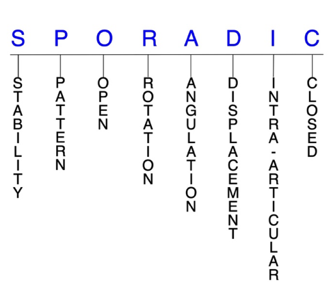
Hand Surgery Resource’s Fracture Description and Characterization Acronym

SPORADIC

S – Stability; P – Pattern; O – Open; R – Rotation; A – Angulation; D – Displacement; I – Intra-articular; C – Closed


S - Stability (stable or unstable)

  • Universally accepted definitions of clinical fracture stability is not well defined in the hand surgery literature.9-11
  • Stable: fracture fragment pattern is generally nondisplaced or minimally displaced. It does not require reduction, and the fracture fragment’s alignment is maintained with simple splinting. However, most definitions define a stable fracture as one that will maintain anatomical alignment after a simple closed reduction and splinting. Some authors add that stable fractures remain aligned, even when adjacent joints are put to a partial range of motion (ROM).
  • Unstable: will notremain anatomically or nearly anatomically aligned after a successful closed reduction and simple splinting. Typically unstable hook of hamate fractures are very rare but painful nonunions are common.  Hamate body fractures are frequently unstable secondary to the extensor carpi ulnaris (ECU) and flexor carpi ulnaris (FCU) attachments on the adjacent pisiform and the fifth metacarpal base.12This is particularly true for fractures in the coronal plane, including the so-called dorsal flake fracture, as they are frequently associated with subluxation or dislocation of the ring and little carpometacarpal (CMC) joints. Fracture-dislocations that involve >33% of the CMC articular surface are most likely to be unstable.2,5,12

P - Pattern

  • Hamate hook: may occur in the distal tip, medial section, or base7 Hook of hamate fractures are typically classified as type 1.6 Fractures of the hook of hamate tip are usually avulsion fractures and have been found to account for ~11% of these injuries.13 Hook of hamate fractures of the medial third—or waist—are also rare, and account for ~13% of hook of hamate fractures.6,13 Base fractures are by far the most common, representing ~76% of hook of hamate fractures.13 Hook of hamate fractures most commonly occur in sports that involve racquets, bats, or clubs, through direct trauma or indirect mechanisms.
  • During a forceful swing, the base of the racquet, bat, or club can impinge against the hook of the hamate and cause a fracture by transmitting the force of the swing through it.4
  • Hamate body: may be oblique, transverse, coronal, or comminuted.3-5,14 Typically classified as type 2 and further divided into two major groups: coronal (type 2A) and transverse (type 2B) fractures
  • Type 2A fractures can be further classified as dorsal oblique or splitting fractures, and these injuries are commonly associated with CMC dislocations.
  • Type 2B fractures are frequently associated with axial ulnar disruptions.4,14 Others have suggested classifying hamate body fractures into the following 4 major groups: sagittal oblique fractures, dorsal coronal fractures, proximal pole fractures, and fractures of the medial tuberosity
  • The position of the wrist at the moment of axial injury to the carpus partially determines the morphology of hamate body fractures in this classification system:
    • With the wrist in ulnar deviation, abutment of the proximal hamate on the triquetrum results in a coronal fracture.
    • When a similar axial load is applied with the wrist in radial deviation or a neutral position, the hamate fractures in the sagittal plane.
    • Proximal pole fractures are typically articular osteochondral shear injuries occurring in association with complex carpal fracture-dislocations.
    • Medial tuberosity fractures characteristically occur following blunt trauma to the ulnar aspect of the carpus.5
    • Combined fractures involving the body and hook of hamate are rare.6

O - Open

  • Open fractures of the hamate are very rare.  In an open hamate fracture, a wound connects the external environment to the fracture site. The wound provides a pathway for bacteria to reach and infect the fracture site. As a result, there is always a risk for chronic osteomyelitis. Therefore, open fractures of the hamate require antibiotics with surgical irrigation and wound debridement.9,15,16
  • Open fractures of the hamate may also require surgical exploration to determine if articular surfaces are involved. After irrigation and debridement, these wounds are occasionally left open and further treatment is typically delayed until the wound shows no sign of infection.17,18

R - Rotation

  • Hamate fracture deformity can be caused by rotation of the distal fragment on the proximal fragment.
  • Degree of malrotation of the fracture fragments can be used to describe the fracture deformity.

A - Angulation (fracture fragments in relationship to one another)

  • Angulation is measured in degrees after identifying the direction of the apex of the angulation.
  • Straight: no angulatory deformity
  • Angulated: bent at the fracture site
  • In small carpal bones like the hamate, angulation of the fracture fragments can be difficult to assess.

D - Displacement (Contour)

  • Displaced: disrupted cortical contours
  • Nondisplaced: fracture line defining one or several fracture fragments; however, the external cortical contours are not significantly disrupted
  • In general, it has been remarked that if displacement does occur in hamate body fractures, it is not usually severe due to the presence of the strong intercarpal ligaments.The displacement seen in distal dorsal hamate fractures associated with CMC subluxation may require surgical attention.
  • A clear definition for displacement in hook of hamate fracture has not yet been established in the literature, but according to some criteria, 30-32% of these fractures are displaced.3,20
  • Due to its anatomic proximity to several ligaments, tendons, and nerves, an acute or chronically nonunited displaced hook of hamate fracture may impinge on the adjacent branch of the ulnar nerve or tendons.21
  • Coronal fractures through the dorsal surface of the hamate body are often associated with subluxation or dislocation of the CMC joint caused by traction of ECU and hypothenar muscles with proximal displacement of the hamate fragment and ring and little metacarpals.2,19

I - Intra-articular involvement

  • Fractures that enter a joint with one or more of their fracture lines.
  • A hamate body fracture can have fragment involvement with any of its CMC or intercarpal joint articulations.
  • If a fracture line enters a joint but does not displace the articular surface of the joint, then it is unlikely that this fracture will predispose to posttraumatic osteoarthritis. If the articular surface is separated or there is a step-off in the articular surface, then the congruity of the joint will be compromised and the risk of posttraumatic osteoarthritis increases significantly.
  • Coronal fractures of the hamate body have also been referred to as intra-articular hamate fractures; however, these injuries are not to be confused with intra-articular fractures of the little metacarpal base.5,19 Hamate proximal pole fractures are typically articular osteochondral shear injuries that are associated with complex carpal fracture-dislocations.5

C - Closed 

  • Closed: no associated wounds; the external environment has no connection to the fracture site or any of the fracture fragments.9-11

Hamate fractures: named fractures, fractures with eponyms and other special fractures

Hamate dislocation and fracture-dislocation

  • Hamate dislocations may occur in isolation, but are more commonly associated with other complex fractures and/or dislocations of the carpus or metacarpals.5 Isolated hamate dislocations are rare and may be volar or dorsal, and there is usually painful swelling of the hand and a bony prominence, along with shortening of the carpus on the ulnar side.22
  • One of the most common fracture patterns is a hamate body fracture with a fracture-dislocation of the ring and little CMC joints, with or without ring and little metacarpal base fractures.12 These hamatometacarpal fracture-dislocations are usually sustained when a clenched fist strikes an unyielding object, and the point of impact on the two metacarpals—as well as the degree of CMC flexion during load transmission—determines the extent of CMC joint injury. The ECU tendon and hypothenar musculature are considered to be the major deforming forces.5
    • Some believe that hamatometacarpalfracture-dislocations are similar enough to coronal fractures of the hamate body that the terms can be used interchangeably, while others consider them to be separate entities. Regardless of the terminology used, it appears that these injuries can be appropriately grouped together.5,19
    • Hamatometacarpal fracture-dislocations are often classified into the following 3 groups:
      • Type IA: subluxation or dislocation of the little CMC joint without a hamate avulsion fracture
      • Type IB: identical to type IA lesions but also include a small dorsal rim hamate avulsion fracture
      • Type II: subluxation or dislocation of the little CMC joint and comminution of the dorsal hamate rim
      • Type III: coronal splitting fractures of the hamate body; depending on the degree and direction of displacement, these injuries may be associated with instability of the ring and little CMC joints2,5,19

Imaging

  • Lateral radiographic views are helpful for showing subluxation or dislocation at the ring and little CMC joints, while oblique views are useful for identifying hamate dorsal avulsion fractures. Anteroposterior (AP) and posteroanterior (PA) views are not very effective for visualizing these injuries.1,3
  • Special X-ray views, such as ulnar-deviated PA, carpal tunnel, and clenched fist views, may also be needed to properly visualize hamatometacarpal fracture-dislocations.5
  • CT scans and MRIs may be used when the diagnosis remains in question or when radiographs raise suspicion for a hamate fracture.1,4,12

Treatment

  • Some authors recommend conservative treatment consisting of closed reduction and immobilization for stable type IA and IB cases, especially if there does not seem to be any risk of recurrent instability after the reduction.2,5 Failure to achieve a reduction of the dislocation in these cases with the corresponding CMC joint congruence may lead to a reduction of grip strength.2

The ring and little CMC joints are quite mobile, with as much as 30° arc of flexion and extension possible at the little CMC joint and ~15° at the ring CMC. Because of this mobility and the instability associated with hamatometacarpal fracture-dislocations, surgical treatment is recommended if displacement or subluxation of the joint is evident, or when closed reduction fails.4  Surgical options include closed reduction and percutaneous pin fixation (CRPP) with K-wires, open reduction and internal fixation (ORIF), and primary excision.2,5  In types II and III injuries, ORIF is indicated to reconstruct soft tissues and restore and fix the hamate bone fragment using a plate or screws to create joint congruence. K-wires may also be considered if the stability of the CMC joint is in doubt.2

Complications

  • Infection
  • Impaired wrist ROM 
  • Reduced wrist strength
  • Nonunion
  • Ulnar nerve palsy

Outcomes

  • Restoration of the articular surface and congruent reduction of the 2 CMC joints increases the chances of achieving an optimal functional outcome.5
  • In one study in which both the ring and little CMC joints were dislocated, fixation of the coronal hamate fracture with screws and temporary bridge plate fixation of the ring CMC joint—when there was CMC instability despite hamate fixation—always led to spontaneous and stable reduction of the little CMC joint because of the ligamentous attachments, and 9 of 11 patients had full recovery.4

Related Anatomy

  • The hamate is an irregularly shaped bone roughly resembling a triangle that is located in the most ulnar position of the distal carpal row. It consists of a wedge-shaped body that articulates distally with the ring and little metacarpals at their respective CMC joints, radially with the capitate, medially with the triquetrum, and proximally with the lunate, as well as a hamular process—or hook. The hook of hamate is a long, thin, curved osseous prominence located on the lower, ulnar-medial surface that projects from the hamate body into the palm in a volar fashion, 1-2 cm distal and lateral to the pisiform in the hypothenar eminence. It also contributes to the ulnar border of the carpal tunnel and the radial border of Guyon’s canal.2,4-6,8
  • Ligamentous attachments of the hamate include the flexor retinaculum, which attaches to the apex of the hook of hamate, the transverse carpal ligament, pisohamate ligament, triquetrohamate ligament, and capitohamate ligament.2,23
  • Tendons associated with the hamate include the flexor digiti minimi brevis and opponens digiti minimi tendons, both of which arise from the convex surface of the hook of hamate, the abductor digiti minimi tendon arising from the hook of hamate, the FCU tendon, which inserts via the pisohamate ligament onto the apex of the hook of hamate, and the flexor digitorum profundus tendon, which uses the hook of hamate as a pulley during flexion of the little finger with ulnar deviation.2,4,6,21
  • Ulnar nerve and motor branch.

Incidence and Related injuries/conditions

  • Fractures of the carpal bones have been found to account for 8-18% of all hand fractures24,25and ~6% of fractures overall.26
  • Fractures of the proximal carpals are more common than the distal carpals, and the most commonly fractured carpal bone is the scaphoid, which represents 58-89% of all carpal fractures.24,25,27,28
  • Fractures of the other 7 carpals are very rare and only account for ~1.1% of all fractures. The triquetrum ranks highest of these bones, while fractures of the remaining carpals are even less common and vary in incidence.29-31
  • Most reports cite the incidence of hamate fractures—including the body and hook—as 2-4% of all carpal fractures;32,33however, it is commonly agreed that many cases of hamate fractures are missed or misdiagnosed, and their true incidence may actually be much higher, with some citing this figure to be 7%25or 15% of all carpal fractures.34
  • Males are much more likely than females to sustain a hamate body fracture.14
  • The specific incidence of hook of hamate fractures is also not well defined, but it is generally agreed that they occur more frequently than body fractures.7,34 The majority of hook fractures (~76%) occur at its base, followed by the medial third (~13%) and distal third (~11%).6,13 The incidence of hook of hamate fractures among professional and recreational athletes in golf, racquet sports, and baseball is much higher than in the general population.6 Only 19.4% of all hook fractures are diagnosed early and 30-32% of them are displaced.20

Work-up Options

  • Routine X-rays
    • Hamate fractures are generally difficult to detect on routine radiographs, but most agree that hamate body fractures are easier to visualize.  However, plain radiographs have low sensitivity and specificity for hook of hamate fractures.1,3,5,7
    • The main radiological characteristics of dorsal hamate body fractures include a 5-10-mm oblong fragment of bone, which projects immediately over the dorsal surface of the hamate.1,3
  • This fragment is most visible on the pronation oblique and/or lateral projections and is not seen on the posteroanterior (PA) projections.1,3
  • Oblique views are also important for identifying hamate dorsal avulsion fractures.12
  • The degree of pronation for oblique views recommended by different investigators has ranged from 15-40°, with less obliquity showing the more radial hamate-ring metacarpal articulation.
  • A lateral radiograph shows subluxation or dislocation at the ring and little CMC joints.5
  • On PA radiographs, the hook of hamate overlaps with the midportion of the distal hamate body and projects as an ovoid bony density, and radiographic signs of this fracture include an absent or indistinct hook or sclerosis of the hook.6
  • Disappearance of the hook on a PA view—due to congenital absence, hypoplasia, surgical resection of the hook, or erosion by amyloid tumor of the carpal tunnel—can mimic a hook of hamate fracture.3
  • Special X-ray views:  Ulnar-deviated PA, carpal tunnel, clenched fist, semi-supinated, and lateral with thumb abduction and radial deviation views may be helpful for assessing both hamate body and hook fractures and associated soft tissue and neural elements.5-7,12
  • The carpal tunnel view cannot always be obtained in the acute phase of injury because it requires hyperextension of the wrist, which can be limited by pain and swelling.3
  • Fractures at the midpoint or distal tip of the hamate hook are more readily detected in special views, whereas the more common fractures at the base are more commonly obscured.3
  • CT scan:  Indicated when the diagnosis remains in question, when an accurate visualization of the carpus is sought, or once radiographs raise suspicion for hamate fracture for more complete fracture characterization, since it provides the most information on the position of the fracture fragment, its possible displacement, and the amount of articular surface involved.1,3,4,12 Considered the radiographic technique-of-choice for suspected acute or chronic bony abnormalities that may suggest a hook of hamate fracture, as it is highly sensitive and specific, with an accuracy of 97.2%.6,7
  • Axial 2-mm cuts through the wrist should be obtained with the hands in the prayer position. This axis is parallel to the long axis of the hook of the hamate and clearly demonstrates the fracture and its displacement, while also allowing comparison between the 2 wrists.3
  • Also useful for providing information on the degree of fracture displacement.6
  • MRI:  An alternative to CT scans for radiographically occult pathologic conditions involving the hamate.MRI is less accurate than CT scans for visualizing hook cortical fractures, but superior for characterizing associated bone marrow edema, ulnar nerve injury, tendon abnormalities, and carpal tunnel abnormalities.  Can be performed instead of a CT scan if the patient lacks neurologic and/or vascular competency in order to better view soft tissue structures.3
  • Bone scan
  • Hamate body and hook fractures are often missed or misdiagnosed as wrist sprains at emergency department presentation, which can lead to severe wrist dysfunction. This highlights the need for clinical suspicion of hamate fractures and careful evaluation of wrist injuries.4,12
ICD-10 Codes

HAMATE & HOOK OF HAMATE FRACTURES

Diagnostic Guide Name

HAMATE & HOOK OF HAMATE FRACTURES

ICD 10 Diagnosis, Single Code, Left Code, Right Code and Bilateral Code

DIAGNOSIS SINGLE CODE ONLY LEFT RIGHT BILATERAL (If Available)
FRACTURE CARPAL BONE - HAMATE        
- DISPLACED   S62.142_ S62.141  
- NONDISPLACED   S62.145_ S62.144_  
FRACTURE CARPAL BONE - HOOK OF HAMATE        
- DISPLACED   S62.155_ S62.154_  
- NONDISPLACED        

Instructions (ICD 10 CM 2020, U.S. Version)

THE APPROPRIATE SEVENTH CHARACTER IS TO BE ADDED TO EACH CODE FROM CATEGORY S62
  Closed Fractures Open Type I or II or Other Open Type IIIA, IIIB, or IIIC
Initial Encounter A B C
Subsequent Routine Healing D E F
Subsequent Delayed Healing G H J
Subsequent Nonunion K M N
Subsequent Malunion P Q R
Sequela S S S

ICD-10 Reference

Reproduced from the International statistical classification of diseases and related health problems, 10th revision, Fifth edition, 2016. Geneva, World Health Organization, 2016 https://apps.who.int/iris/handle/10665/246208

Clinical Presentation Photos and Related Diagrams
Hamate and Hook of Hamate Fracture Diagrams
  • Hamate Dorsal Lip Fracture (curved arrow) nondisplaced
    Hamate Dorsal Lip Fracture (curved arrow) nondisplaced
  • Hamate Dorsal Lip Fracture (curved arrow) displaced
    Hamate Dorsal Lip Fracture (curved arrow) displaced
  • Hook of Hamate Fracture (curved arrow) nondisplaced; 1-Trapezial ridge, 2-carpal tunnel, 3-Hook of Hamate, 4-Pisiform; Dotted line = transverse carpal ligament
    Hook of Hamate Fracture (curved arrow) nondisplaced; 1-Trapezial ridge, 2-carpal tunnel, 3-Hook of Hamate, 4-Pisiform; Dotted line = transverse carpal ligament
Symptoms
History of trauma
Fracture pain and deformity
Fracture deformity, swelling, crepitus, ecchymosis
Impaired grip strength
Ulnar nerve paresthesia and carpal tunnel syndrome symptoms
Abnormal bony prominence and/or loss of knuckle prominence when associated with ring or little CMC joint subluxations
Typical History

A typical patient is a 20-year old, left-handed collegiate male golfer who injured his wrist during a recent tournament match. While swinging at a ball that was positioned off the fairway near a tree, he missed the shot and connected with a large tree root in the vicinity. This force twisted the butt of the club against the hook of hamate in his right wrist, sending most of its energy directly to the hook and fracturing it. The trauma caused immediate pain, swelling, and tenderness in the man’s wrist, which led him to remove himself from the match and seek medical attention.

Positive Tests, Exams or Signs
Work-up Options
Images (X-Ray, MRI, etc.)
Hamate Fracture Imaging
  • Routine AP Wrist X-ray does not demonstrate the hook of hamate fracture.
    Routine AP Wrist X-ray does not demonstrate the hook of hamate fracture.
  • CT Scan sagittal images do demonstrate the hook of hamate fracture (arrow)
    CT Scan sagittal images do demonstrate the hook of hamate fracture (arrow)
  • Bone Scan shows marked increased up-take at the site of the hook of hamate fracture  (arrow)
    Bone Scan shows marked increased up-take at the site of the hook of hamate fracture (arrow)
Treatment Options
Treatment Goals
  • There are 4 basic goals when treating closed hamate fractures:9,16
    1. A wrist with a normal appearance. The X-ray may not need to be perfect, but the hamate should have no obvious deformity (ie, the hamate looks normal!)
    2. Avoid stiffness by maintaining a normal functional ROM (ie, the wrist works!)
    3. The hamate is not painful (ie, the hamate does not hurt!)
    4. Congruent joint surface with none-to-minimal joint surface irregularities (ie, the joints associated with the hamate do not develop early post-traumatic arthritis!)
    5. Fracture care for open fractures should minimize the risk for infection and osteomyelitis.
Conservative

Hamate body

  • The main goal of treatment for hamate body fractures is to preserve a stable distal hamate articular surface, as well as ring and little CMC joint function.9
  • Conservative treatment consisting of closed reduction and immobilization with a cast or aluminum, plaster, fiberglass, or custom splint may be recommended for stable, isolated hamate body fractures that are nondisplaced or minimally displaced and those that involve <33% of the CMC articulation.1,11,12,18 This type of conservative approach is particularly recommended for stable type 2A fractures, but these instances are rare since most coronal hamate body fractures are unstable.14,35  This wrist should be casted in mid dorsiflexion.
  • Closed reduction of coronal dorsal oblique fractures is relatively simple, but may be more difficult with coronal splitting fractures.14 Frequent imaging should be used during the immobilization period to monitor for loss of reduction and improper healing.12 Although isolated hamate body fractures do occur, they are more commonly part of a larger injury pattern and surgical treatment is often required.1
  • Even hamate body fractures that require a reduction to correct fracture-related deformity usually can be held in anatomic or near-anatomic alignment with a splint without internal or external surgical fixation.18

Hook of hamate

  • Managing hook of hamate fractures—especially those older than 3 months—is controversial, and there are no established standards, but most experts agree that treatment should be contingent on the acuity of the diagnosis, degree of displacement, and location of the fracture.6,7
  • Many clinicians believe that conservative treatment is likely to result in nonunion and therefore advocate surgery for all patients, but many others continue to recommend a trial of immobilization.7
    • Patients with nondisplaced hook of hamate base fractures without comminution that present within 3 months of injury are the best candidates to be managed conservatively.4-6,36 Conservative treatment typically consists of immobilization using a cast or aluminum, plaster, fiberglass, or custom splint.1,6,10,11  Casting should maintain the transverse palmar arches and keep the wrist neutral for mild dorsiflexion.
  • Cast immobilization has generally been found to provide consistent healing if treatment is started within 7 days of the fracture, but overall healing rates have been identified as ~50% for all hook of hamate fractures, with waist and tip fractures having even lower union rates due to poor vascularity.4,36 Despite this, distal third hook fractures are most likely to receive conservative treatment regardless of other factors. Low-intensity pulsed ultrasound has also been advanced as an alternative conservative intervention for non-displaced hook of hamate fractures.6
Operative
  • Surgical treatment of hamate fractures must always be an individualized therapeutic decision. However, surgical hamate fracture care is most frequently recommended when:
    1. Closed reduction fails or the simple splint or cast immobilization does not maintain the reduction. For these irreducible or unstable fractures, operative treatment is recommended to achieve the 4 treatment goals of fracture care.
    2. When there is a significantly displaced hamate body fracture fragment involving one of its associated joints. Surgical fracture care is often required.
    3. Open hamate fractures require surgical care in the form of irrigation and debridement to prevent chronic infection.

Hamate body

  • CRPP is recommended for most unstable type 2A fractures.  It may also be utilized for some type 2B fractures, where it has been found to achieve bony union in most cases.14
  • ORIF is typically needed for hamate body fractures involving more than one-third of the CMC articulation surfaces and cases in which accurate and stable reduction is not achieved percutaneously.14 A dorsal approach is the most common approach.  The optimal method for internal fixation is controversial, as K-wires, Herbert screws, and cortical mini-lag screws have all been utilized, sometimes in conjunction.19
  • Cortical screws are usually needed to fixate large hamate fragments.12 In addition to ORIF with either compression screws or low-profile plates, these injuries may also require temporary fixation across the CMC joints to aid in stability.37

Hook of hamate

  • In addition to the criteria listed above, surgery is commonly recommended for symptomatic displaced hook of hamate fractures, those that are >3 months old, partial or complete nonunion, ulnar or median nerve or ulnar artery compression, tendon rupture, metacarpal subluxation, patients who are unable to keep the affected wrist immobilized, and recurrent cases.3,4
  • The 2 most common surgical procedures for hook of hamate fractures are surgical hook excision and ORIF.3,4
    • Surgical hook excision
      • More commonly performed and generally regarded as the gold standard for acute displaced hook fractures and nonunions.5,7
      • Some authors recommend early excision over ORIF for hook fractures that are displaced, chronic, or present with ulnar nerve compression, because multiple studies have shown no adverse sequelae on wrist ROM or grip strength with it.36
      • Involves a curvilinear, volar carpal tunnel approach, followed by a careful dissection of the hook, flexor tendons and ulnar nerve and artery. Careful dissection is essential to avoid complications such as an injury to the deep motor branch of the ulnar nerve.   The ulnar motor branches leave the ulnar nerve in a straight dorsal direction before turning radial.  As the ulnar motor branch goes dorsal, it travels against the distal surface of the hook before turning further radial under the small fat pad on the roof of the carpal tunnel.  The hook is then excised at the base with a sharp osteotome, and afterwards, the ligament attachments to the tip of the hook are repaired. The hand is then immobilized in a short arm cast for 4-6 weeks.3,4
    • ORIF
      • Operative techniques include open volar screw or K-wire fixation, hook plate fixation, and dorsal percutaneous screw fixation.13
      • The rationale for using this procedure on a hook of hamate fracture or nonunion may be based on an estimated 11% loss of flexor tendon excursion after excision of the hook of hamate. Another advantage is that it generally provides good symptom relief and preserves the hook as a pulley.4,3 The importance of preserving the hook of the hamate as a pulley for the medial flexor tendons is another matter that continues to be debated.3,4
Treatment Photos and Diagrams
Surgical Treatment of Hook of Hamate Fractures
  • Carpal tunnel incision for excision of the hook of the hamate. Incision will be opened from distal to proxima,l starting at the distal end of the carpal tunnel and Guyon's Canal.  The transverse carpal ligament will be incised proximally until the radial aspect of the hamate is exposed.  Guyon's canal will be opened with transectiion of the pisohamate ligament and exposure of the ulnar nerve and artery.
    Carpal tunnel incision for excision of the hook of the hamate. Incision will be opened from distal to proxima,l starting at the distal end of the carpal tunnel and Guyon's Canal. The transverse carpal ligament will be incised proximally until the radial aspect of the hamate is exposed. Guyon's canal will be opened with transectiion of the pisohamate ligament and exposure of the ulnar nerve and artery.
  • Initial dissection and initial exposure of the hook of the hamate has been done.  The ulnar neurovascular bundle (1) is exposed. The periosteum (2) has been dissected off the hook of the hamate (3).  The hypothenar muscle origins (4) have been released from the hook.  The radial periosteum is being stripped of the hook and the flexors tendons (5) are retracted.
    Initial dissection and initial exposure of the hook of the hamate has been done. The ulnar neurovascular bundle (1) is exposed. The periosteum (2) has been dissected off the hook of the hamate (3). The hypothenar muscle origins (4) have been released from the hook. The radial periosteum is being stripped of the hook and the flexors tendons (5) are retracted.
  • Beaver blade removing ulnar periosteum (2) off the hook of the hamate (1).  Ulnar neurovascular bundle (3) visible.
    Beaver blade removing ulnar periosteum (2) off the hook of the hamate (1). Ulnar neurovascular bundle (3) visible.
  • Hook of Hamate (1) exposure complete.  The hypothenar muscle origins (2) have been released from the hook.  The flexors tendons (3) are retracted.
    Hook of Hamate (1) exposure complete. The hypothenar muscle origins (2) have been released from the hook. The flexors tendons (3) are retracted.
CPT Codes for Treatment Options

Per an agreement between Hand Surgery Resource, LLC and the American Medical Association (AMA) users are required to accept the following End User Point and Click Agreement in order to view CPT content on this website.  Please read and then click "Accept" at the bottom to indicate your acceptance of the agreement.

End User Point and Click Agreement

CPT codes, descriptions and other data only are copyright 2019 American Medical Association (AMA). All rights reserved. CPT is a registered trademark of the AMA. The AMA reserves all rights to approve any license with any Federal agency.

You, as an individual, are authorized to use CPT only as contained in Hand Surgery Resource solely for your own personal information and only within the United States for non-commercial, educational use for the purpose of education relating to the fundamental principles of hand surgery and the common diseases, disorders and injuries affecting the human hand. You agree to take all necessary steps to ensure your compliance with the terms of this agreement.

Any use not authorized herein is prohibited, including by way of illustration and not by way of limitation, making copies of CPT for resale and/or license, transferring copies of CPT to any party not bound by this agreement, creating any modified or derivative work of CPT, or making any commercial use of CPT. License to use CPT for any use not authorized herein must be obtained through the AMA, CPT Intellectual Property Services, 330 N. Wabash Avenue, Chicago, IL 60611. Applications are available at the AMA Web site, http://www.ama-assn.org/go/cpt.

Common Procedure Name
Excision trapezium, pisiform, scaphoid of hook of hamate
CPT Description
Carpectomy; one bone
CPT Code Number
25210
CPT Code References

The American Medical Association (AMA) and Hand Surgery Resource, LLC have entered into a royalty free agreement which allows Hand Surgery Resource to provide our users with 75 commonly used hand surgery related CPT Codes for educational promises. For procedures associated with this Diagnostic Guide the CPT Codes are provided above. Reference materials for these codes is provided below. If the CPT Codes for the for the procedures associated with this Diagnostic Guide are not listed, then Hand Surgery Resource recommends using the references below to identify the proper CPT Codes.

 CPT QuickRef App.  For Apple devices: App Store. For Android devices: Google Play

 CPT 2021 Professional Edition: Spiralbound

Hand Therapy

Post-treatment Management

  • The care and precautions related to immobilization devices for the hamate fracture must be carefully reviewed with the patient. Patients should be educated regarding care and precautions. Patients should know that pain, especially increasing pain, numbness, tingling, skin irritation, splint loosening, or excessive splint tightness are red flags and should be reported to the surgeon or his team.
  • Pain should be managed with properly fitting splints, reassurance, elevation, ice in the initial post-fracture period, and mild pain medications. Patients should be encouraged to discontinue pain medication as soon as possible. Opioid use should be kept to a minimum.
  • Patients should be instructed to carefully exercise all joints in the injured hand, wrist, and arm that do not require immobilization.Patients usually can exercise on their own; however, signs of generalized stiffness are indications for referral to hand therapy (PT or OT).
  • Conservative treatment for hamate body and hook fractures usually requires immobilization with casting for ~6 weeks, usually followed by an additional 4-6 weeks of physical therapy.
  • If the injury is treated surgically with ORIF, the patient will typically require casting for 2-3 weeks, followed by an additional 4 weeks of physical therapy without placing strain on the affected wrist, before progressing to full activity in 6-8 weeks.
  • If hook excision is performed, the patient can begin physical therapy in 2-4 weeks without limitations and can return to full activity within 6-8 weeks.  Gentle finger motion can be started early but wrist and power grip exercises should be delayed while the repaired ligaments around the hook heal.
    • Return to full activity is dependent on the initial fracture treatment. Typically, if treated conservatively, simple hamate fractures heal within 6-8 weeks of injury.3
  • If an infection does occur, management should focus on eradicating sepsis with thorough debridement, appropriate antibiotics (eg, cephalosporin, penicillin), and fracture stabilization to obtain fracture union and to regain a functional extremity.17
Complications
  • Complications of inadequate hamate body fracture treatment include fracture malreduction, nonunion, CMC joint posttraumatic arthritis, and chronic pain.  Additional complications of surgical fixation of these injuries include infection and soft tissue scarring.12
  • The most common complication of hook of hamate fractures appears to be nonunion.  This complication is particularly common in patients treated conservatively and has been found to persist even in cases of early diagnosis and immobilization.5
  • Retrospective analyses have demonstrated nonunion rates greater than 50% and as high as 80-90% with conservative treatment.3  Some clinicians believe that nonunion is the natural course of these fractures rather than a complication because of the poor blood supply to the distal fragment, motion at the fracture site, wide displacement of the fragments, and delayed diagnosis.7,39
  • Avascular necrosis is extremely rare in hamate body fractures because of the bone’s 3-vessel vascular supply, but may develop in hook of hamate fractures.12,39
  • Ulnar nerve palsy—which can result from contusion of the nerve or pressure caused by hemorrhage and edema.  It is also possible in hamate body fractures and isolated dorsal dislocations of the base of the ring and the little metacarpals without hamate fracture.23
  • Other possible complications of hamate body and hook fractures include flexor tendon rupture, ulnar nerve deficits, osteonecrosis (AVN) of the hook, ulnar artery compression/thrombosis, chronic hypothenar pain, and carpal tunnel syndrome.3,36
  • In one survey of 133 surgeons who had excised the hamate hook at least once, the overall self-reported complication rate was 3%.  Intraoperative complications included flexor tendon adhesions, division of the motor branch of the ulnar nerve, and division of the superficial palmar arch.  Postoperative complications included transient median and ulnar sensory neuropathy and diminished grip strength, and it is reasonable to assume that similar complications could be expected with ORIF.5
Outcomes
  • When hamate body and hook fractures are diagnosed in a timely manner and managed appropriately, outcomes are generally positive.3,4,7,8,13,14,40 
  • In one study, 14 patients with type 2 fractures (11 type 2A and 3 type 2B) were treated surgically with either CRPP or ORIF.
  • Nondisplaced hamate body fractures treated with cast immobilization for 6 weeks have been found to lead to good healing in patients.3
  • All type 2A patients achieved bony union and regained full ROM of the wrist and fingers. 
  • All type 2B patients achieved bony union, although 2 had ulnar nerve palsy and limited ROM of the wrist and fingers and decreased grip strength; however, all of these patients had experienced severe compression of the wrist between heavy weights that led to soft tissue and nerve damage. The authors therefore concluded that outcomes appear to be based more by associated soft tissue damage than fracture type.14
  • In another study, 14 patients had type 2A fractures sustained primarily from punching injuries, and 12 displayed subluxation of the ring and/or little CMC joints. Treatment was either conservative—using a wrist splint or plaster cast—or operative. Surgical techniques included fixation with either K-wires or compact hand screws alone, or a combination of these techniques.  Conservative treatment led to good functional outcomes in the patients with nondisplaced type 2A fractures.
  • Outcomes for the patients treated surgically were more mixed but generally positive, with some participants experiencing impaired wrist function. The authors concluded that delays in diagnosis and treatment, as well as residual deformities with dorsal subluxation of the metacarpal may be confounding variables associated with poorer outcomes.13
  • Although some authors advocate conservative treatment with lower arm cast immobilization for hook of hamate fractures, failure rates of 90-100% with painful nonunion and other complications have been reported.39  In one review of 24 acute hook fractures treated by lower arm cast immobilization, 11 patients healed successfully, while another 11 required immediate fragment excision, and there were no residual complaints in those who went on to have surgery.20
  • In one study of 21 patients with hook of hamate fractures, 17 were treated with surgical excision, 3 with ORIF, and 1 conservatively.  Of the 19 patients available for follow-up, patient satisfaction was high, with only 1 patient being dissatisfied after failure of ORIF when his painful nonunion persisted. Excision was subsequently required.8
  • In another retrospective study of 51 patients with hook of hamate fractures, 14 were treated surgically and 37 conservatively.   Patient outcomes were good to excellent regardless of the treatment chosen, with minimal pain and good hand functionality.7
  • In the largest single series in the literature, consistently satisfactory results were reported after 59 cases of hook of hamate excision, with normal grip strength being restored in 57 patients by 6 months after surgery.40
Key Educational Points
  • Underlying pathological conditions such as bone tumors—like enchodromas—and osteoporosis should be expected in hamate fractures that occur from trivial trauma.
  • The functional needs of each patient must be considered when recommending treatment for hamate fractures.
  • Even though hamate fractures are uncommon, their incidence seems to be increasing due to the growing popularity of golf and other racquet sports.5
  • Although non-scaphoid carpal bones like the hamate have received considerably less attention than the scaphoid itself, these injuries can still produce morbidity that is disproportionate to their incidence for 2 primary reasons:
  • These fractures may have a subtle clinical and radiographic presentation and are easily overlooked or misdiagnosed, in some cases as wrist sprains. This can lead to suboptimal management and poor long-term outcomes with significant wrist disability.1,41
  • These fractures are often harbingers of significant ligamentous disruption or associated carpal fractures, and failure to recognize a more global injury pattern can result in undertreatment and permanent wrist dysfunction.1
  • Understanding the mechanism and clinical presentation of hamate fractures is the first step to proper diagnosis. Radiologists and acute care clinicians familiar with the subtle radiographic findings can further characterize these fractures with CT evaluation, which aids in hamate fracture diagnosis and characterization, and facilitates orthopaedic management that will optimize long-term patient outcomes.12
  • Because the hook of hamate acts as a fulcrum for the ulnar digital flexors, some surgeons believe that excising it results in a loss of grip strength, particularly when the hand is held in ulnar deviation.38
  • Despite its continued importance as a key anatomic landmark in modern medicine, the hook of the hamate remains a source of diagnostic errors in current clinical practice.6
  • Acute traumatic hook of hamate fractures may present with other fractures of the distal upper extremity, such as distal ulnar fractures, other carpal bone fractures, hamate dislocations, and medial CMC joint disruptions.6
  • Although CT is the most reliable imaging test to visualize and subsequently diagnose hook of hamate fractures, taking CT scans of all wrist injuries is probably not a reasonable solution. Clinicians must therefore work towards establishing more effective criteria for determining when this diagnostic tool is necessary.4
References

New and Cited Articles

  1. Shah MA, Viegas SF. Fractures of the carpal bone excluding the scaphoid. J Hand Surg Am 2002; 2(3): 129-140
  2. Sarabia Condés JM, Ibañez Martínez L, Sánchez Carrasco MA, et al. Hamate fractures. Rev Esp Cir Ortop Traumatol2015;59(5):299-306.PMID: 25823609
  3. Mouzopoulos G, Vlachos C, Karantzalis L, Vlachos K. Fractures of hamate: a clinical overview. Musculoskelet Surg2018. [Epub] PMID: 29845407
  4. Papp S. Carpal bone fractures. Hand Clin2010;26(1):119-27. PMID: 20006250
  5. O'Shea K, Weiland AJ. Fractures of the hamate and pisiform bones. Hand Clin2012;28(3):287-300. PMID: 22883867
  6. Davis DL. Hook of the Hamate: The Spectrum of Often Missed Pathologic Findings. AJR Am J Roentgenol2017;209(5):1110-1118. PMID: 28834449
  7. Kadar A, Bishop AT, Suchyta MA, Moran SL. Diagnosis and management of hook of hamate fractures. J Hand Surg Eur Vol2018;43(5):539-545. PMID: 28893146
  8. Bishop AT, Beckenbaugh RD. Fracture of the hamate hook. J Hand Surg Am1988;13(1):135-9. PMID: 3351218
  9. Cheah AE, Yao J. Hand Fractures: Indications, the Tried and True and New Innovations.J Hand Surg Am 2016;41:712-22. PMID: 27113910
  10. Nesbitt KS, Failla JM, Les C. Assessment of instability factors in adult distal radius fractures. J Hand Surg Am 2004;29:1128-38.PMID: 15576227
  11. Walenkamp MM, Vos LM, Strackee SD, Goslings JC, Schep NW. The Unstable Distal Radius Fracture-How Do We Define It? A Systematic Review. J Wrist Surg 2015;4:307-16. PMID: 26649263
  12. Cecava ND, Finn MF, Mansfield LT. Subtle radiographic signs of hamate body fracture: a diagnosis not to miss in the emergency department. Emerg Radiol2017;24(6):689-695. PMID: 28616787
  13. Wharton DM, Casaletto JA, Choa R, Brown DJ. Outcome following coronal fractures of the hamate. J Hand Surg Eur Vol2010;35(2):146-9. PMID: 19282405
  14. Hirano K, Inoue G. Classification and treatment of hamate fractures. Hand Surg2005;10(2-3):151-7. PMID: 16568508
  15. Ketonis C, Dwyer J, Ilyas AM. Timing of Debridement and Infection Rates in Open Fractures of the Hand: A Systematic Review. Hand (N Y) 2017;12:119-26. PMID: 28344521
  16. Meals C, Meals R. Hand fractures: a review of current treatment strategies. J Hand Surg Am 2013;38:1021-31. PMID: 23618458
  17. Day CS. Fractures of the Metacarpals and Phalanges. In: Green DP, ed. Green's Operative Hand Surgery. Seventh ed. Philadelphia: Elsevier; 2016, pp. 231-77.
  18. Weinstein LP, Hanel DP. Metacarpal fractures.J Hand Surg Am 2002; 2(4):168–180.
  19. Kaneko K, Ono A, Uta S, et al. Hamatometacarpal fracture-dislocation: distinctive three dimensional computed tomographic appearance. Chir Main2002;21(1):41-5. PMID: 11885387
  20. Carroll RE, Lakin JF. Fracture of the hook of the hamate: acute treatment. J Trauma1993;34(6):803-5. PMID: 8315674
  21. Bachoura A, Wroblewski A, Jacoby SM, et al. Hook of hamate fractures in competitive baseball players. Hand (N Y)2013;8(3):302-7. PMID: 24426938
  22. Gunn RS. Dislocation of the hamate bone. J Hand Surg Br1985;10(1):107-8. PMID: 3998586
  23. Fakih RR, Fraser AM, Pimpalnerkar AL. Hamate fracture with dislocation of the ring and little finger metacarpals. J Hand Surg Br1998;23(1):96-7. PMID: 9571493
  24. van Onselen EB, Karim RB, Hage JJ, Ritt MJ. Prevalence and distribution of hand fractures. J Hand Surg Br2003;28(5):491-5. PMID: 12954264
  25. Hove LM. Fractures of the hand. Distribution and relative incidence. Scand J Plast Reconstr Surg Hand Surg1993;27(4):317-9. PMID: 8159947
  26. Dobyns JH, Beckenbaugh RD, Bryan RS, et al. Fractures of the hand and wrist. In: Flynn JE, editor. Hand surgery. Third ed. Philadelphia: Lippincott Williams & Wilkins; 1982.
  27. Rhemrev SJ, Ootes D, Beeres FJ, et al. Current methods of diagnosis and treatment of scaphoid fractures. Int J Emerg Med2011;4:4. PMID: 21408000
  28. Hey HW, Chong AK, Murphy D. Prevalence of carpal fracture in Singapore. J Hand Surg Am2011;36(2):278-83. PMID: 21276891
  29. Larsen CF, Brøndum V, Skov O. Epidemiology of scaphoid fractures in Odense, Denmark. Acta Orthop Scand1992;63(2):216-8. PMID: 1590062
  30. Höcker K, Menschik A. Chip fractures of the triquetrum. Mechanism, classification and results. J Hand Surg Br1994;19(5):584-8. PMID: 7822914
  31. Garcia-Elias M.Dorsal fractures of the triquetrum-avulsion or compression fractures? J Hand Surg Am1987;12(2):266-8. PMID: 3559084
  32. Roche S, Lenehan B, Street J, O’Sullivan M. Fourth metacarpal base fracture in association with coronal hamate fracture. Injury Extra 2005;36: 316–8.
  33. Geissler WB. Carpal fractures in athletes. Clin Sports Med2001;20(1):167-88. PMID: 11227704
  34. Welling RD, Jacobson JA, Jamadar DA, et al. MDCT and radiography of wrist fractures: radiographic sensitivity and fracture patterns. AJR Am J Roentgenol2008;190(1):10-6. PMID: 18094287
  35. Cano Gala C, Pescador Hernández D, Rendón Díaz DA, et al. Fracture of the body of hamate associated with a fracture of the base of fourth metacarpal: A case report and review of literature of the last 20 years. Int J Surg Case Rep2013;4(5):442-5. PMID: 23557937
  36. Suh N, Ek ET, Wolfe SW. Carpal fractures. J Hand Surg Am2014;39(4):785-91. PMID: 24679911
  37. Snoap T, Habeck J, Ruiter T. Hamate Fracture. Eplasty2015;15:ic28. eCollection 2015. PMID: 26171103
  38. Klausmeyer MA, Mudgal CS. Hook of hamate fractures. J Hand Surg Am2013;38(12):2457-60. PMID: 23891177
  39. Scheufler O, Andresen R, Radmer S, et al. Hook of hamate fractures: critical evaluation of different therapeutic procedures. Plast Reconstr Surg2005;115(2):488-97. PMID: 15692355
  40. Stark HH, Chao EK, Zemel NP, et al. Fracture of the hook of the hamate. J Bone Joint Surg Am1989;71(8):1202-7. PMID: 2777848
  41. Zoltie N. Fractures of the body of the hamate. Injury1991;22(6):459-62. PMID: 1757137
  42. Brach P, Goitz R. An update on the management of carpal fractures. J Hand Ther2003;16(2):152-60. PMID: 12755166
  43. Pan T, Lögters TT, Windolf J, Kaufmann R. Uncommon carpal fractures. Eur J Trauma Emerg Surg2016;42(1):15-27. PMID: 26676306

Classics

  1. Bowen TL. Injuries of the hamate bone. Hand1973;5(3):235-8. PMID: 4747858
  2. Nisenfield FG, Neviaser RJ. Fracture of the hook of the hamate: a diagnosis easily missed. J Trauma1974;14(7):612-6. PMID: 4841621