Hand Surgery Source

FRACTURE DISTAL RADIUS SALTER I-V PEDIATRIC

Introduction

Fracture Nomenclature for Pediatric Distal Radius Salter I-V Fractures

Hand Surgery Resource’s Diagnostic Guides describe fractures by the anatomical name of the fractured bone and then characterize the fracture by the Acronym:

In addition, anatomically named fractures are often also identified by specific eponyms or other special features.

For the Pediatric Distal Radius Salter I-V Fracture, the historical and specifically named fractures include:

None

By selecting the name (diagnosis), you will be linked to the introduction section of this Diagnostic Guide dedicated to the selected fracture eponym.


Although the bones of adults and children share many of the same risks for fracture, children are uniquely susceptible to physeal fracture, as their bones are still growing, and the growth plate is the weakest part of the growing bone. Growth plates are located at the ends of the long bones, and they help determine the ultimate length and shape of mature bones. Thus, these fractures require prompt attention. If not treated properly, an arm or leg could grow to be crooked or of unequal length compared to the other limb. In 1963, Salter and Harris proposed a classification system for pediatric physeal fractures. The Type II fracture is the most common type of physeal fracture. In the Salter/Harris classification, the higher the number, the more likely the growth plate will be permanently damaged and bone deformity will occur.

Definitions

  • A pediatric distal radius fracture is a disruption of the mechanical integrity of the distal radius.
  • A pediatric distal radius fracture produces a discontinuity in the distal radius contours that can be complete or incomplete.
  • A pediatric distal radius fracture is caused by a direct force that exceeds the breaking point of the bone.

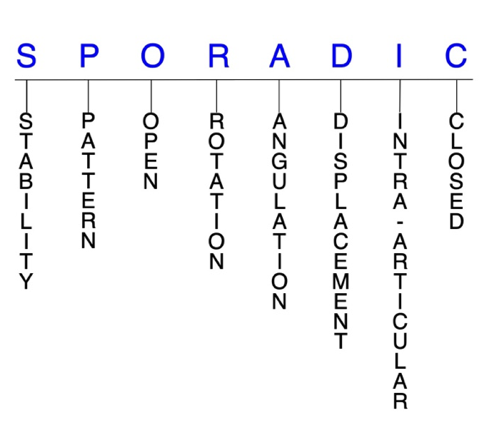
Hand Surgery Resource’s Fracture Description and Characterization Acronym

SPORADIC

S – Stability; P – Pattern; O – Open; R – Rotation; A – Angulation; D – Displacement; I – Intra-articular; C – Closed


S - Stability (stable or unstable)

  • Universally accepted definitions of clinical fracture stability is not well defined in the literature.1-3
  • Stable: fracture fragment pattern is generally nondisplaced or minimally displaced. It does not require reduction, and the fracture fragments’ alignment is maintained by with simple splinting or casting. However, most definitions define a stable fracture as one that will maintain anatomical alignment after a simple closed reduction and splinting. Some authors add that stable fractures remain aligned, even when adjacent joints are put to a partial range of motion (ROM).
  • Unstable: will not remain anatomically or nearly anatomically aligned after a successful closed reduction and immobilization. Typically, unstable pediatric distal radius fractures have significant deformity with comminution, displacement, angulation, and/or shortening.
    • A pediatric distal radius fracture is further defined as unstable if it is unable to resist displacement after being anatomically reduced.4

P - Pattern

  • Distal radial styloid
  • Distal dorsal medial fragment
  • Distal volar medial fragment
  • Distal radial shaft

O - Open

  • Open: a wound connects the external environment to the fracture site. The wound provides a pathway for bacteria to reach and infect the fracture site. As a result, there is always a risk for chronic osteomyelitis. Therefore, open fractures of the pediatric distal radius require antibiotics with surgical irrigation and wound debridement.1,5,6

R - Rotation

  • Pediatric distal radius fracture deformity can be caused by proximal rotation of the fracture fragment in relation to the distal fracture fragment.
  • Degree of malrotation of the fracture fragments can be used to describe the fracture deformity.

A - Angulation (fracture fragments in relationship to one another)

  • Angulation is measured in degrees after identifying the direction of the apex of the angulation.
  • Straight: no angulatory deformity
  • Angulated: bent at the fracture site
  • Extreme dorsal angulation may be associated with triangular fibrocartilage complex (TFCC) injuries.4

D - Displacement (Contour)

  • Displaced: disrupted cortical contours
  • Nondisplaced: ≥1 fracture lines defining one or several fracture fragments; however, the external cortical contours are not significantly disrupted
  • In pediatric distal radius fractures, displacement can be either extra-articular or intra-articular. Extra-articular displacement can occur in any of the 3 planes. When displacement occurs in the sagittal plane, it typically leads to loss of the palmar tilt, while volar shear injuries tend to increase palmar tilt. In the coronal plane, displacement typically manifests as the loss of radial inclination and/or height.7

I - Intra-articular involvement

  • Intra-articular fractures are those that enter a joint with ≥1 of their fracture lines.
  • Distal radius fractures can have fragment involvement with the radiocarpal joint or distal radioulnar joint (DRUJ).
  • If a fracture line enters a joint but does not displace the articular surface of the joint, then it is unlikely that this fracture will predispose to post-traumatic osteoarthritis. If the articular surface is separated or there is a step-off in the articular surface, then the congruity of the joint will be compromised and the risk of post-traumatic osteoarthritis increases significantly.

C - Closed

  • Closed: no associated wounds; the external environment has no connection to the fracture site or any of the fracture fragments.4-6

Related Anatomy8,9

  • The radius consists of a radial head and radial neck at its proximal end. The shaft of the radius then extends from the neck and has a rectangular epiphysis on its distal end. The articular—or lateral—surface of the distal radius is biconcave and triangular, and the apex of this triangle is directed toward the styloid process. On the medial surface, there is a concavity called the ulnar notch that articulates with the head of ulna, forming the DRUJ. The distal surface of the radius has two facets for articulation with the scaphoid and lunate carpal bones. There is also an articulation between the distal radius and the triquetral bone facilitated by a biconcave articular disc. Collectively, these three articulations form the radiocarpal joint.
  • Ligaments associated with the distal radius include the dorsal radiocarpal ligament, which spans the ulnar aspect of the dorsal rim of the distal radius from the ulnar margin of the Lister tubercle to the sigmoid notch; the radioscaphocapitate ligament, which originates from the radial styloid and spans to the volar rim of the distal radius at the scaphoid fossa; the long radiolunate ligament, which originates from the volar rim of the scaphoid fossa; and the short radiolunate ligament, which originates from the volar rim of the lunate fossa.
  • Tendons associated with the distal radius include those associated with the extensor carpi radialis brevis (ECRB), extensor carpi radialis longus (ECRL), and EPL muscles. The dorsal tubercle protrudes on the posterior aspect of the distal head of the radius and is seated between the grooves for the tendons of the ECRB and ECRL as well as the tendon of the EPL.
  • The radius and ulna are connected by a sheet of thick fibrous tissue called the interosseous membrane.

Incidence

  • Salter-Harris type II fractures occur after age 10 years.
  • Physeal fractures are twice as likely in boys as in girls, since girls finish growing earlier than boys (14 vs 16 years).
  • Approximately 33% of all growth plate fractures occur during competitive sports (e.g., football, basketball, gymnastics), while about 20% occur during recreational sporting activities (e.g., biking, skiing, skateboarding).
ICD-10 Codes

FRACTURE DISTAL RADIUS PEDIATRIC

Diagnostic Guide Name

FRACTURE DISTAL RADIUS PEDIATRIC

ICD 10 Diagnosis, Single Code, Left Code, Right Code and Bilateral Code

DIAGNOSIS SINGLE CODE ONLY LEFT RIGHT BILATERAL (If Available)
FRACTURE DISTAL RADIUS - PHYSEAL    /td>    
- SALTER HARRIS TYPE I   S59.212_ S59.211_  
- SALTER HARRIS TYPE II   S59.222_ S59.221_  
- SALTER HARRIS TYPE III   S59.232_ S59.231_  
- SALTER HARRIS TYPE IV   S59.242_ S59.241_  

Instructions (ICD 10 CM 2020, U.S. Version)

THE APPROPRIATE SEVENTH CHARACTER IS TO BE ADDED TO EACH CODE FROM CATEGORY S59.2
  Closed Fractures
Initial Encounter A
Subsequent Routine Healing D
Subsequent Delayed Healing G
Subsequent Nonunion K
Subsequent Malunion P
Sequela S

ICD-10 Reference

Reproduced from the International statistical classification of diseases and related health problems, 10th revision, Fifth edition, 2016. Geneva, World Health Organization, 2016 https://apps.who.int/iris/handle/10665/246208

Clinical Presentation Photos and Related Diagrams
  • Silver Fork Deformity from Salter II Distal Radius Fracture
    Silver Fork Deformity from Salter II Distal Radius Fracture
Basic Science Photos and Related Diagrams
  • Normal Growth Plate  A = diaphysis; B = metaphysis; C = growth plate (epiphyseal plate); D = epiphysis
    Normal Growth Plate A = diaphysis; B = metaphysis; C = growth plate (epiphyseal plate); D = epiphysis
  • Salter I Fracture - Fracture line through growth plate (physis) but non displaced
    Salter I Fracture - Fracture line through growth plate (physis) but non displaced
  • Salter II Fracture - Fracture line through growth plate (physis) and metaphysis. The metaphyseal fragment is called the Thurston-Holland fragment.
    Salter II Fracture - Fracture line through growth plate (physis) and metaphysis. The metaphyseal fragment is called the Thurston-Holland fragment.
  • Salter III Fracture - Fracture line through growth plate (physis) and the epiphysis and articular surface.
    Salter III Fracture - Fracture line through growth plate (physis) and the epiphysis and articular surface.
  • Salter IV Fracture - A vertical fracture line through metaphysis, the growth plate (physis) and the epiphysis and articular surface.
    Salter IV Fracture - A vertical fracture line through metaphysis, the growth plate (physis) and the epiphysis and articular surface.
  • Salter V Fracture - A compression-type fracture line in the physis with extension in multiple directions. Growth plate (physis) damage is likely.
    Salter V Fracture - A compression-type fracture line in the physis with extension in multiple directions. Growth plate (physis) damage is likely.
  • Histology of the normal Epiphysis, Growth Plate and Metaphysis:  A = Articular cartilage; B = Subchondral bone; C = Cancellous bone; D = Medullary cavity; E = Cancellous bone; F = Resting cartilage; G = Proliferating cartilage; H = Hypertrophy; I – Calcification; J = Vascular invasion
    Histology of the normal Epiphysis, Growth Plate and Metaphysis: A = Articular cartilage; B = Subchondral bone; C = Cancellous bone; D = Medullary cavity; E = Cancellous bone; F = Resting cartilage; G = Proliferating cartilage; H = Hypertrophy; I – Calcification; J = Vascular invasion
Symptoms
History of trauma - fall on the outstretched hand (FOOSH)
Pain, swelling and bruising of the wrist
Pain with wrist motion or pressure on the wrist area
Wrist deformity (silver fork deformity)
Patient is a growing child
Typical History
  • A junior high school soccer player fell over a teammate during a scrimmage and landed on her outstretched left hand and wrist.  She is complaining of pain, swelling and deformity in the right wrist.  The team’s athletic trainer has applied a wrist splint and ice pack while the patient waits for a ride to the local emergency room.
Positive Tests, Exams or Signs
Work-up Options
Images (X-Ray, MRI, etc.)
  • Salter II Fracture Distal Radius Lateral View
    Salter II Fracture Distal Radius Lateral View
  • Salter II Fracture Distal Radius AP View
    Salter II Fracture Distal Radius AP View
Treatment Options
Treatment Goals
  • When treating closed pediatric distal radius fractures, the treating surgeon has 4 basic goals:1,6
    1. A wrist with a normal appearance. The X-ray may not need to be perfect, but the wrist should have no obvious deformity (i.e., the wrist looks normal!)
    2. Avoid wrist stiffness by maintaining a normal functional ROM (ie, the wrist works!)
    3. The wrist is not painful (i.e., the wrist does not hurt!)
    4. Congruent joint surface with none-to-minimal joint surface irregularities (ie, the joint does not develop early post-traumatic arthritis!)
  • One additional goal is mandatory for open fractures:
    1. Fracture care should minimize the risk for infection and osteomyelitis.
Conservative
  • The typical closed, nondisplaced, minimally angulated, pediatric distal radius fracture without significant malrotation can be managed conservatively.15
  • For most stable distal radius fractures, closed reduction and immobilization is the preferred treatment approach. After the fracture is reduced, a splint may be used within the first few days to account for swelling. Next, the wrist is to be immobilized by a plaster cast or removable splint.14
Operative
  • Surgical treatment of pediatric distal radius fractures must always be an individualized therapeutic decision.8,14,15  However, surgical distal radius fracture care is most frequently recommended when:
    1. Closed reduction fails or the simple splint or cast immobilization does not maintain the reduction. For these irreducible or unstable fractures, operative treatment is recommended to achieve the 4 treatment goals of fracture care.
    2. There is a significantly displaced distal radius fracture involving the DRUJ or radiocarpal joint.
    3. Open distal radius fractures. These injuries require surgical care in the form of irrigation and debridement to prevent chronic infection.
  • When surgery is indicated, the surgeon must select from numerous fixation options, and their decision should evaluate for the biomechanical characteristics of each method, the associated difficulty and advantages/disadvantages of the procedure, and any soft tissue morbidity present.14
  • Closed reduction and internal fixation
    • Most suitable procedure for fractures without articular involvement or substantial metaphyseal comminution.14
    • Intramedullary nailing or K-wire pinning can be used for fixation.
  • Open reduction and internal fixation
    • Dorsal plates
    • Volar plates
  • Fragment-specific fixation
  • Arthroscopy
  • The care and precautions related to immobilization devices for the pediatric distal radius fracture must be carefully reviewed with the patient. Patients should be educated regarding care and precautions. Patients should know that pain, especially increasing pain, numbness, tingling, skin irritation, splint loosening, or excessive tightness are red flags and should be reported to the surgeon or his team.
  • Pain should be managed with properly fitting splints and casts, reassurance, elevation, ice in the initial post-fracture period, and mild pain medications. Patients should be encouraged to discontinue pain medication as soon as possible. Opioid use should be kept to a minimum.
  • Joints that are splinted for closed stable fractures are usually immobilized.
  • Fractures that require internal fixation can be mobilized after 4 weeks.
  • Home exercise programs are typically recommended over formal supervised postoperative rehabilitation, as the two approaches generally lead to comparable outcomes.13
Complications
  • Deformity
  • Bony bridge across fracture line that stunts bone growth or causes bone to angulate, grows more slowly than normal, or stops growing completely at the injured site
  • Some fractures stimulate growth, causing bones to become longer than the opposite limb
  • Malunion
  • Post-traumatic osteoarthritis
  • Osteomyelitis
  • Carpal malalignment
  • TFCC tears
  • Carpal tunnel syndrome
  • Forearm compartment syndrome
  • Non-union
  • Stiffness
Outcomes
  • Growth plate fractures must be watched carefully to ensure good long-term results.
  • More complicated fractures may require follow-up visits until the child reaches skeletal maturity.
  • Parents must be informed that this is a growth-related fracture, and it may affect growth even with proper care.
  • The rate of healing is relatively high in patients with open physes, and fractures closer to the distal physes have greater remodeling potential than those closer to the proximal physes.
  • Although many surgical techniques are available for these injuries, there are certain limitations with each type of fixation. For example, fixation with plate and screws produces an excellent and stable reduction but results in large unaesthetic incisions and slower healing.
  • Satisfactory outcomes are likely with closed reduction and casting when the fracture is held in acceptable alignment.
Key Educational Points
  • Pediatric distal radius Salter-Harris I-V fractures occur much more frequently in boys than girls, and the age of maximal incidence is somewhat younger for girls than for boys. This is thought to be associated with the greater exposure of boys to trauma and to the relative delay of epiphyseal closure in boys rather than to any intrinsic difference in epiphyseal structure between the sexes.
  • Most pediatric distal radius fractures were traditionally treated conservatively, but the paradigm has shifted more recently towards a surgical approach based on new research findings and the development of newer surgical instruments and techniques.
  • The fracture line is at the junction of the bone and the epiphyseal plate. Most pediatric distal radius Salter-Harris I-V fractures are produced by a shearing force, and there is not much damage to the epiphyseal plate cells, therefore, growth is not disturbed.
  • Ability to remodel residual angulation after fracture depends on the age of the patient at the time of fracture, the remaining time and the distance between the fracture and the epiphyseal plate, and the extent of residual angulation following reduction.
  • The potential for remodeling is maximal when the plane of deformity lies in the plane of motion of the adjacent joint.
  • Children under 10 years old possess the ability to correct angulation up to 28°; the potential for correction is decreased with greater angulation and increasing age beyond 10 years of age. Therefore, it is recommended that correction of angular deformities should be performed in children over 10–12 years.
  • There is a correlation between premature closure of the growth plate and multiple and/or forceful attempts at closed reduction.
  • Radiology studies - X-ray13,14
    • Plain radiographs are the gold standard in diagnosing pediatric distal radius fractures.
    • Standard views
      1. Posteroanterior view
      2. Oblique view
      3. Lateral view
    • Special views
      1. 10 tilt lateral view
      2. Lateral view with beam inclined 20°
      3. 45° pronated oblique view
  • Radiology studies - Computerized tomography (CT) scanning
    • Can be valuable to ensure acceptable articular alignment when conservative treatment is being considered and during surgical planning for complex fracture patterns.12,14
  • Magnetic resonance imaging - MRI without contrast
    • Reserved for cases in which it is unclear whether a fracture is truly present, and to identify concomitant soft tissue pathology.14
References

Cited Articles

  1. Cheah, AE and Yao, J. Hand Fractures: Indications, the Tried and True and New Innovations. J Hand Surg Am 2016;41(6):712-22. PMID: 27113910
  2. Nesbitt, KS, Failla, JM and Les, C. Assessment of instability factors in adult distal radius fractures. J Hand Surg Am 2004;29(6):1128-38. PMID: 15576227
  3. Walenkamp, MM, Vos, LM, Strackee, SD, et al. The Unstable Distal Radius Fracture-How Do We Define It? A Systematic Review. J Wrist Surg 2015;4(4):307-16. PMID: 26649263
  4. Porrino, JA, Jr., Maloney, E, Scherer, K, et al. Fracture of the distal radius: epidemiology and premanagement radiographic characterization. AJR Am J Roentgenol 2014;203(3):551-9. PMID: 25148157
  5. Ketonis, C, Dwyer, J and Ilyas, AM. Timing of Debridement and Infection Rates in Open Fractures of the Hand: A Systematic Review. Hand (N Y) 2017;12(2):119-126. PMID: 28344521
  6. Meals, C and Meals, R. Hand fractures: a review of current treatment strategies. J Hand Surg Am 2013;38(5):1021-31. PMID: 23618458
  7. Katt, B, Seigerman, D, Lutsky, K, et al. Distal Radius Malunion. J Hand Surg Am 2020;45(5):433-442. PMID: 32220492
  8. Fernandez, DL and Jupiter, JB. Fractures of the Distal Radius: A Practical Approch to Management. 2002;2nd edition.
  9. Zumstein, MA, Hasan, AP, McGuire, DT, et al. Distal radius attachments of the radiocarpal ligaments: an anatomical study. J Wrist Surg 2013;2(4):346-50. PMID: 24436840
  10. Richard, MJ, Katolik, LI, Hanel, DP, et al. Distraction plating for the treatment of highly comminuted distal radius fractures in elderly patients. J Hand Surg Am 2012;37(5):948-56. PMID: 22480509
  11. Meena, S, Sharma, P, Sambharia, AK, et al. Fractures of distal radius: an overview. J Family Med Prim Care 2014;3(4):325-32. PMID: 25657938
  12. Alluri, RK, Hill, JR and Ghiassi, A. Distal Radius Fractures: Approaches, Indications, and Techniques. J Hand Surg Am 2016;41(8):845-54. PMID: 27342171
  13. Brogan, DM, Richard, MJ, Ruch, D, et al. Management of Severely Comminuted Distal Radius Fractures. J Hand Surg Am 2015;40(9):1905-14. PMID: 26243322
  14. Schneppendahl, J, Windolf, J and Kaufmann, RA. Distal radius fractures: current concepts. J Hand Surg Am 2012;37(8):1718-25. PMID: 22763062
  15. Bales, JG and Stern, PJ. Treatment strategies of distal radius fractures. Hand Clin 2012;28(2):177-84. PMID: 22554661
  16. Kreder, HJ, Hanel, DP, Agel, J, et al. Indirect reduction and percutaneous fixation versus open reduction and internal fixation for displaced intra-articular fractures of the distal radius: a randomised, controlled trial. J Bone Joint Surg Br 2005;87(6):829-36. PMID: 15911668

New Articles

  1. Sferopoulos NK. Classification of distal radius physeal fractures not included in the salter-harris system. Open Orthop J 2014;8:219-24. PMID: 25132871
  2. Lollino N, et al. Salter-Harris type II proximal humerus injuries: our experience with a new external fixator. Tech Hand Up Extrem Surg 2013;17(3):176-8. PMID: 23970202
  3. Waters PM, Bae DS, Montgomery KD. Surgical management of post-traumatic distal radial growth arrest in adolescents. J Pediatr Orthop 2002;22(6):717- 724. PMID: 12409894
  4. Elia G, Blood T, Got C. The Management of Pediatric Open Forearm Fractures. J Hand Surg Am 2020;45(6):523-527. PMID: 32265052
  5. Passiatore M, De Vitis R, Perna A, et al. Extraphyseal distal radius fracture in children: is the cast always needed? A retrospective analysis comparing Epibloc system and K-wire pinning. Eur J Orthop Surg Traumatol2020;30(7):1243-1250. PMID: 32405758
  6. Georgiadis AG, Burgess JK, Truong WH, Janicki JA. Displaced Distal Radius Fracture Treatment: A Survey of POSNA Membership. J Pediatr Orthop 2020;40(9):e827-e832. PMID: 32271318
  7. Rabinovich RV, Shore BJ, Glotzbecker M, et al. The Effect of Casting Simulation on Maintenance of Fracture Alignment Following Closed Reduction of Pediatric Distal Radius Fractures: Does More Simulation Matter? J Surg Educ 2021;S1931-7204(21)00056-8. PMID: 33896733

Reviews

  1. Verdano MA, et al. Salter-Harris type II proximal humerus injuries: state-of-the-art treatment. Musculoskelet Surg2012;96(3):155-9. PMID: 22879059
  2. Brown JH, DeLuca SA. Growth plate injuries: Salter-Harris classification. Am Fam Physician 1992;46(4):1180-4. PMID: 1414883

Classics

  1. Lesko PD, Georgis T, Slabaugh P. Irreducible Salter-Harris type II fracture of the distal radial epiphysis. J Pediatr Orthop 1987;7(6):719-21. PMID: 3429661
  2. Salter RB, Harris WR. Injuries involving the epiphyseal plate. J Bone Joint Surg 1963;45A:587-622.