Hand Surgery Source

FRACTURE RADIUS AND/OR ULNA AND/OR BOTH FOREARM BONES

Introduction

Fracture Nomenclature for Fracture Radius and/or Ulna and/or Both Forearm Bones

Hand Surgery Resource’s Diagnostic Guides describe fractures by the anatomical name of the fractured bone and then characterize the fracture by the Acronym:

In addition, anatomically named fractures are often also identified by specific eponyms or other special features.

For the Fracture Radius and/or Ulna and/or Both Forearm Bones, the historical and specifically named fractures include:

Galeazzi fracture

Monteggia fracture

By selecting the name (diagnosis), you will be linked to the introduction section of this Diagnostic Guide dedicated to the selected fracture eponym.


Radial and ulnar shaft fractures are relatively common injuries. Although either the radial shaft or ulnar shaft can be fractured in isolation, in most cases both bones are injured simultaneously, and these are referred to as “both bone forearm fractures.” There is a bimodal age distribution for forearm fractures, with one peak in teenage male patients and another in women over the age of 60 years. In adults, these injuries are typically due to motor vehicle accidents, athletic injuries, and falls from height, while low-energy trauma can also produce a fracture in individuals with impaired bone quality. Although conservative treatment may be indicated in certain cases, the majority of forearm fractures are managed surgically through open reduction and internal fixation (ORIF).1-4

Definitions

  • Forearm fractures are a disruption of the mechanical integrity of the radial and/or ulnar shaft.
  • Forearm fractures produce a discontinuity in the radial and/or ulnar shaft contours that can be complete or incomplete.
  • Forearm fractures are caused by a direct force that exceeds the breaking point of the bone.

Hand Surgery Resource’s Fracture Description and Characterization Acronym
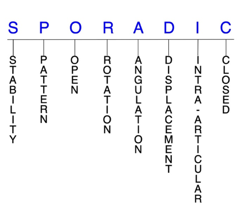
SPORADIC

S – Stability; P – Pattern; O – Open; R – Rotation; A – Angulation; D – Displacement; I – Intra-articular; C – Closed


S - Stability (stable or unstable)

  • Universally accepted definitions of clinical fracture stability are not well defined in the literature.5-7
  • Stable: fracture fragment pattern is generally nondisplaced or minimally displaced. It does not require reduction, and the fracture fragments’ alignment is maintained with simple splinting or casting. However, most definitions define a stable fracture as one that will maintain anatomical alignment after a simple closed reduction and splinting. Some authors add that stable fractures remain aligned, even when adjacent joints are put to a partial range of motion (ROM).
  • Unstable: will not remain anatomically or nearly anatomically aligned after a successful closed reduction and immobilization. Typically unstable radial and ulnar shaft fractures have significant deformity with comminution, displacement, angulation, and/or shortening.
  • For radial shaft fractures, the likelihood of instability is higher when the injury site is more distal than proximal.8
  • An unstable ulnar shaft fracture is one that involves >50% displacement, >10° angulation, the proximal third of the bone, or which also features proximal radioulnar joint (PRUJ) or distal radioulnar joint (DRUJ) instability. Most ulnar shaft fractures, however, are stable.2

P - Pattern

  • Radial shaft
    • Proximal third
    • Middle third
    • Distal third
    • Apex volar
    • Apex dorsal
  • Ulnar shaft
    • Proximal third
    • Middle third
    • Distal third
    • Apex volar
    • Apex dorsal

O - Open

  • Open: a wound connects the external environment to the fracture site. The wound provides a pathway for bacteria to reach and infect the fracture site. As a result, there is always a risk for chronic osteomyelitis. Therefore, open forearm fractures require antibiotics with surgical irrigation and wound debridement.5,9,10
  • There is high risk for visible deformity and open injury in both bone forearm fractures due to the significant forces involved.4
  • Many isolated ulnar shaft fractures are also open, while open radial shaft fractures are not uncommon.1,2

R - Rotation

  • Forearm fracture deformity can be caused by proximal rotation of the fracture fragment in relation to the distal fracture fragment. Rotational deformities are common after forearm fractures.
  • Degree of malrotation of the fracture fragments can be used to describe the fracture deformity.

A - Angulation (fracture fragments in relationship to one another)

  • Angulation is measured in degrees after identifying the direction of the apex of the angulation.
  • Straight: no angulatory deformity
  • Angulated: bent at the fracture site
  • Galeazzi fractures have been found to result in shortening and angulation, which is responsible for the disruption of the DRUJ.8

D - Displacement (contour)

  • Displaced: disrupted cortical contours
  • Nondisplaced: ≥1 fracture lines defining one or several fracture fragments; however, the external cortical contours are not significantly disrupted

I - Intra-articular involvement

  • Intra-articular fractures are those that enter a joint with ≥1 of their fracture lines.
  • Forearm fractures can have fragment involvement with the DRUJ, PRUJ, radiocarpal, humeroulnar, or humeroradial joints.
  • If a fracture line enters a joint but does not displace the articular surface of the joint, then it is unlikely that this fracture will predispose to post-traumatic osteoarthritis. If the articular surface is separated or there is a step-off in the articular surface, then the congruity of the joint will be compromised and the risk of post-traumatic osteoarthritis increases significantly.

C - Closed

  • Closed: no associated wounds; the external environment has no connection to the fracture site or any of the fracture fragments.4-6

Forearm fractures: named fractures, fractures with eponyms and other special fractures

Galeazzi fracture

  • Also known as a reverse Monteggia fracture or Piedmont fracture, the Galeazzi fracture is fracture of the middle to distal third of the radial shaft combined with a subluxation or dislocation of the DRUJ.8,11
  • The Galeazzi fracture is considered a true forearm axis injury because concomitant bone and soft tissue injuries—particularly the triangular fibrocartilage complex (TFCC) and/or interosseous membrane (IOM) which contribute to forearm instability.1,8
  • These injuries typically result from direct impact to the radius with forearm pronation. When a patient sustains a radial shaft fracture in the middle to distal third of the bone, suspicion should be raised for an associated DRUJ injury.1,8
  • Galeazzi and Monteggia fractures—discussed next—are more common in pediatric than adult patients.4

Imaging11

  • Radiology studies - X-ray
    • Anteroposterior (AP) and lateral views are usually sufficient, but an oblique view may help to better classify the injury.
    • If a coexistent injury is suspected, the distal wrist and proximal elbow should also be radiographed.
  • Radiology studies - Computerized tomography (CT) scanning
    • Not usually needed, but occasionally used to evaluate for nonunion.
  • Magnetic resonance imaging - MRI without contrast
    • Often used to detect TFCC tears and/or IOM disruption.

Treatment

Conservative

  • Conservative treatment is not recommended for most adults with Galeazzi fractures; however, temporary immobilization with a sugar-tong splint may be used while a patient is awaiting an orthopedic consult or surgery.11

Operative

  • Surgery is required in the majority of cases and first involves rigid fixation of the radius. This is followed by intraoperative assessment of DRUJ stability.
    • If the DRUJ is stable after fixation. This may be followed by immobilization in supination or a neutral position for 2-4 weeks.
    • If the DRUJ is unstable, the TFCC may be repaired and/or the forearm pinned in supination in a reduced position.8
    • If an ulnar styloid fracture accompanies the unstable DRUJ, the styloid should be repaired with fixation using a lag screw, tension band wire or with excision of the fragment and repair of the TFCC to the fovea.8,11
  • Volar plate fixation
    • Most common surgical approach
    • Fixation may be more difficult when the fracture is in the distal third of the radius, where it may only be possible to insert two screws distal to the fracture line in these cases if using a 3.5-mm straight plate.1 Modern T-plates provide better fixation in the distal fragment by allowing additional screws in the distal fragment.
  • Open reduction of the DRUJ11
    • Sometimes necessary when the DRUJ cannot be reduced closed.
    • The TFCC should be repaired and/or the forearm pinned in supination.
    • Followed by immobilization with a splint or cast and the elbow in supination.

Complications

  • Nonunion
  • Malunion
  • DRUJ instability
  • Reduced grip strength

Outcomes

  • Due to their complexity, Galeazzi fractures can lead to a variety of outcomes, and complication rates as high as 40% have been reported.11
  • Closed reduction and immobilization alone has been associated with poor outcomes in >90% of adult patients.11

Monteggia fracture

  • A Monteggia fracture involves a fracture of the proximal third of the ulna combined with a subluxation or dislocation of the radial head at the PRUJ and the humeroradial joint.4,8
  • These injuries most commonly occur secondary to a direct blow to the posterior aspect of the ulna, with the elbow extended and the forearm in hyperpronation.12,13
  • Monteggia fractures account for <2% of all forearm fractures.12

Imaging13

  • Radiology studies
ICD-10 Codes

FRACTURE RADIUS AND ULNAR SHAFTS

Diagnostic Guide Name

FRACTURE RADIUS AND ULNAR SHAFTS

ICD 10 Diagnosis, Single Code, Left Code, Right Code and Bilateral Code

DIAGNOSIS SINGLE CODE ONLY LEFT RIGHT BILATERAL (IF AVAILABLE)
FRACTURE ULNA SHAFT        
- TRANSVERSE         
 - DISPLACED   S52.222_ S52.221_  
 - NONDISPLACED   S52.225_ S52.224_  
- OBLIQUE        
 - DISPLACED   S52.232_ S52.231_  
 - NONDISPLACED   S52.235_ S52.234_  
- COMMINUTED        
 - DISPLACED   S52.252_ S52.251_  
 - NONDISPLACED   S52.255_ S52.254_  
- SPIRAL        
 - DISPLACED   S52.242_ S52.241_  
 - NONDISPLACED   S52.245_ S52.244_  
- GREENSTICK   S52.212_ S52.211_  
- SEGMENTAL        
 - DISPLACED   S52.262_ S52.261_  
 - NONDISPLACED   S52.265_ S52.264_  
- MONTEGGIA'S   S52.272_ S52.271_  
FRACTURE RADIUS SHAFT        
- TRANSVERSE         
 - DISPLACED   S52.322_ S52.321_  
 - NONDISPLCED   S52.325_ S52.324_  
- OBLIQUE        
 - DISPLACED   S52.332_ S52.331_  
 - NONDISPLACED   S52.335_ S52.334_  
- COMMINUTED        
 - DISPLACED   S52.352_ S52.351_  
 - NONDISPLACED   S52.355_ S52.354_  
- SPIRAL        
 - DISPLACED   S52.342_ S52.341_  
 - NONDISPLACED   S52.345_ S52.344_  
- GREENSTICK   S52.312_ S52.311_  
- SEGMENTAL        
 - DISPLACED   S52.362_ S52.361_  
 - NONDISPLACED   S52.365_ S52.364_  
- GALEAZZI'S   S52.372_ S52.371_  

Instructions (ICD 10 CM 2020, U.S. Version)

USE TWO DIAGNOSIS CODES - ONE FOR EACH BONE
THE APPROPRIATE SEVENTH CHARACTER IS TO BE ADDED TO EACH CODE FROM CATEGORY S52
  Closed Fractures Open Type I or II or Other Open Type IIIA, IIIB, or IIIC
Initial Encounter A B C
Subsequent Routine Healing D E F
Subsequent Delayed Healing G H J
Subsequent Nonunion K M N
Subsequent Malunion P Q R
Sequela S S S

ICD-10 Reference

Reproduced from the International statistical classification of diseases and related health problems, 10th revision, Fifth edition, 2016. Geneva, World Health Organization, 2016 https://apps.who.int/iris/handle/10665/246208

Clinical Presentation Photos and Related Diagrams
Closed Radius and Ulna Fractures (Double Bone Forearm Fracture)
  • Left closed mid-third transverse radius and ulna fractures
    Left closed mid-third transverse radius and ulna fractures
Right open distal -third transverse radius and ulna fractures
  • Right open distal -third transverse radius and ulna fractures with transverse wound over the radius.
    Right open distal -third transverse radius and ulna fractures with transverse wound over the radius.
  • Lateral X-ray of right open distal -third transverse radius and ulna fractures
    Lateral X-ray of right open distal -third transverse radius and ulna fractures
  • AP X-ray of right open distal -third transverse radius and ulna fractures
    AP X-ray of right open distal -third transverse radius and ulna fractures
Symptoms
History of trauma
Fracture pain and deformity
Swelling, ecchymosis, and tenderness at fracture site
Typical History

A typical patient is a 67-year-old woman who was injured in a car accident. The woman was traveling as a passenger in a car traveling at a high speed on the highway when the driver failed to notice an upcoming traffic jam and slammed on the brakes, leading to a crash with the stopped car in front of them. The passenger extended both hands forward to brace for impact, slamming them into the front dashboard, and this force led to a fracture of both the right radial and ulnar shafts.

Positive Tests, Exams or Signs
Work-up Options
Images (X-Ray, MRI, etc.)
Radius and Ulna Fractures (Double Bone Forearm Fracture)
  • Left closed mid-third transverse radius and ulna fractures
    Left closed mid-third transverse radius and ulna fractures
  • Severely traumatized right forearm (AP X-ray) with open double bone forearm fractures.
    Severely traumatized right forearm (AP X-ray) with open double bone forearm fractures.
  • Severely traumatized right forearm (Oblique) X-ray) with open double bone forearm fractures.
    Severely traumatized right forearm (Oblique) X-ray) with open double bone forearm fractures.
Treatment Options
Treatment Goals

When treating closed adult double bone forearm fractures, the treating surgeon has 4 basic goals:5,10

  1. A hand with a normal appearance. The X-ray may not need to be perfect, but the forearm should have no obvious deformity (i.e., the forearm looks normal!)
  2. Avoid stiffness by maintaining a normal functional ROM (i.e., the forearm works!)
  3. The forearm is not painful (i.e., the forearm does not hurt!)
  4. If the forearm fracture involves any joint, then a congruent joint surface with none-to-minimal joint surface irregularities (i.e., the wrist, radioulnar and the elbow joints do not develop early post-traumatic arthritis!)
  5. For all open fractures, irrigation and debridement should be done to minimize the risk of infection and osteomyelitis.
Conservative
  • Most forearm fractures will require surgical intervention, but there is a certain set of cases that can be treated conservatively.4
  • Conservative treatment through immobilization is indicated for truly isolated ulnar shaft fractures—the nightstick fracture—especially those that are closed, stable, nondisplaced or minimally displaced (<50%), and located within the middle third of the shaft. Note that most isolated ulnar shaft fractures are stable.1-3
    • Immobilization involves functional casting or short-arm casting, preferably distal to the elbow, in either pronation or supination. Early mobilization is recommended for minimally displaced fractures following removal of the immobilization device. Mid-pronation positioning is also recommended.2
    • Regular follow-up radiographs and clinical examinations are necessary after this approach.4
  • Isolated, nondisplaced radial shaft fractures may also be considered for conservative treatment; however, these fractures are usually displaced, and most cases will still require surgery because closed reduction of the radial bow is challenging and concomitant injury is common.1
  • Most both bone forearm fractures also necessitate surgical intervention, but initial reduction and immobilization is often used to prepare the patient for surgery and to immobilize the injured forearm.4
Operative

Operative1,3,4

  • Surgical treatment of forearm fractures must always be an individualized therapeutic decision. However, surgical forearm fracture care is most frequently recommended when:
  1. Closed reduction fails or the immobilization device does not maintain the reduction. For these irreducible or unstable fractures, operative treatment is recommended to achieve the 4 treatment goals of fracture care.
  2. There is a significantly displaced forearm fracture involving any of the associated joints.
  3. Open forearm fractures which require surgical ORIF and irrigation and debridement to prevent chronic infection.
  • In addition to the indications above, most isolated radial shaft fractures, most both bone forearm fractures, and many proximal ulnar shaft fractures will require surgical intervention.4
  • ORIF
    • ORIF with plate and screw construct is generally accepted as the gold standard for treatment for radial and/or ulnar shaft fractures.
    • Radial shaft
      • Anterior approach or “volar approach of Henry”
        • Indicated for any radial shaft fracture from the proximal neck and bicipital tuberosity through the distal styloid region because it exposes the entire radius. This is the preferred approach for most radial shaft fractures, as disadvantages are minimal.
      • Posterior approach or “Thompson’s approach”
        • Used less frequently because it is less extensile than the volar approach, but it may be appropriate in certain situations.
        • Primary advantage is that it can be used for proximal fractures, especially if the soft tissue of the anterior forearm is compromised. The posterior approach also allows the posterior interosseous nerve to be definitively identified and protected during fixation.
    • Ulnar shaft
      • A direct ulnar approach is typically recommended at the interval between the flexor carpi ulnaris and the extensor carpi ulnaris. A plate is used for fixation which can be placed either dorsal or volar on the ulnar shaft.
    • Both bones
      • Similar surgical methods are used for both bone forearm fractures. The ulnar shaft fracture is typically fixated first through a direct ulnar approach, followed by repair of the radial shaft fracture, usually through the standard volar Henry approach.
  • Intramedullary nailing
    • An alternative surgical approach
  • External fixation which is usually used in severe injuries prior to definitive ORIF.
Treatment Photos and Diagrams
Closed Double Bone Forearm Fracture Treatment
  • Left closed mid-third transverse radius and ulna fracture ORIF with 6 hole plate on radius and 7 hole plate on the ulna.
    Left closed mid-third transverse radius and ulna fracture ORIF with 6 hole plate on radius and 7 hole plate on the ulna.
Double Bone Forearm Fracture Treatment Plate
  • Standard 6 hole plate and self-tapping screws
    Standard 6 hole plate and self-tapping screws
Open Double Bone Forearm Fracture Treatment
  • Right open distal -third transverse radius and ulna fractures with transverse wound over the radius.
    Right open distal -third transverse radius and ulna fractures with transverse wound over the radius.
  • Lateral X-ray right open distal -third transverse radius and ulna fractures
    Lateral X-ray right open distal -third transverse radius and ulna fractures
  • AP X-ray right open distal -third transverse radius and ulna fractures
    AP X-ray right open distal -third transverse radius and ulna fractures
  • Right radial plate and compressed fracture line.
    Right radial plate and compressed fracture line.
  • Right ulna plate and compressed fracture line.
    Right ulna plate and compressed fracture line.
  • Wound after I&D and ORIF of both fractures.  Median nerve visible.
    Wound after I&D and ORIF of both fractures. Median nerve visible.
  • AP X-ray right distal -third transverse radius and ulna fractures after ORIF.
    AP X-ray right distal -third transverse radius and ulna fractures after ORIF.
Healed Radius and Ulna Fractures after ORIF.
  • Healed Radius and Ulna Fractures after ORIF with 8 and 7 hole plates
    Healed Radius and Ulna Fractures after ORIF with 8 and 7 hole plates
Complications after Radius and Ulna Fractures ORIF
  • Broken plate secondary to non-union of the ulna
    Broken plate secondary to non-union of the ulna
  • Right Radius and Ulna Synostosis
    Right Radius and Ulna Synostosis
Hand Therapy

Post-treatment Management

  • The care and precautions related to immobilization devices for forearm fractures must be carefully reviewed with the patient. Patients should be educated regarding care and precautions. Patients should know that pain, especially increasing pain, numbness, tingling, skin irritation, splint loosening, or excessive tightness are red flags and should be quickly reported to the surgeon or his team.
  • Pain should be managed with properly fitting splints and casts, reassurance, elevation, ice in the initial post-fracture period, and mild pain medications. Patients should be encouraged to discontinue pain medication as soon as possible. Opioid use should be kept to a minimum.
  • Joints that are splinted for closed stable fractures are usually immobilized.
  • After surgical fixation of both bone fractures, patients are placed in a splint that immobilizes the elbow and forearm, while the fingers and thumb should be left free to encourage ROM and to prevent stiffness. Patients can usually begin elbow ROM exercises within 5-7 days after surgery. Forearm immobilization should be used after ORIF until the fractures show signs of healing. Most activities can resume after union has been confirmed, which typically takes 2-3 months.4
Complications
  • Nonunion
    • Occurs after 2-10% of all forearm fractures, but rates are generally lower in isolated ulnar shaft fractures.3,19
  • Post-traumatic osteoarthritis
    • Can occur in any of the associated wrist or elbow joints after certain forearm fractures.
  • Chronic osteomyelitis
    • Rare but can occur in open forearm fractures, especially in patients with diabetes or in patients whose immune system is compromised.
  • Malunion
  • Cross-union
  • Radioulnar synostosis
  • TFCC tears
  • Forearm compartment syndrome
  • Infection
  • Neurovascular deficits
  • Stiffness
  • Loss of forearm rotation
Outcomes
  • Literature comparing outcomes between conservative and surgical approaches for forearm fractures is limited.4
  • Most patients with uncomplicated radial shaft fractures can expect to experience positive outcomes, with fractures generally healing functionally within 8 weeks and union rates of up to 95% being reported.1
  • Both surgical and nonsurgical treatment of isolated ulnar shaft fractures have been associated with high union rates and good functional outcomes, especially in cases treated with ORIF and below-elbow brace immobilization, with literature suggesting union rates as high as 100%.2
  • The prognosis for both bone forearm fractures is generally very good as well, with research showing very high rates of union within two months of surgery and only mild impairments being reported.4
Key Educational Points
  • Forearm fractures must be immobilized before radiographic fracture healing is complete to avoid complications.
  • Underlying pathological conditions such as bone tumor and osteoporosis should be expected in fractures that occur from trivial trauma.
  • The functional needs of each patient must be considered when recommending treatment for forearm fractures.
  • Although there are some indications for nonsurgical treatment of forearm fractures, many challenges are associated with this approach, and a significant share of these injuries are therefore treated surgically.1,4
  • For ulnar and radial shaft fractures—open and closed—separate approaches to each bone should be used to reduce the risk synostosis.1
References

Cited Articles

  1. Means, KR, Jr. and Graham, TJ. Chapter 23: Disorders of the Forearm Axis. In Green DP, ed. Green’s Operative Hand Surgery. Sixth ed. Philadelphia: Elsevier;2011, pp. 837-868.
  2. Sauder, DJ and Athwal, GS. Management of isolated ulnar shaft fractures. Hand Clin 2007;23(2):179-84, vi. PMID: 17548009
  3. Davis, DD and Kane, SM. Nightstick Fracture. In: StatPearls. Treasure Island (FL): 2020. PMID: 32310411
  4. Small, RF and Yaish, AM. Radius and Ulnar Shaft Fractures. In: StatPearls. Treasure Island (FL): 2020. PMID: 32491613
  5. Cheah, AE and Yao, J. Hand Fractures: Indications, the Tried and True and New Innovations. J Hand Surg Am 2016;41(6):712-22. PMID: 27113910
  6. Nesbitt, KS, Failla, JM and Les, C. Assessment of instability factors in adult distal radius fractures. J Hand Surg Am 2004;29(6):1128-38. PMID: 15576227
  7. Walenkamp, MM, Vos, LM, Strackee, SD, et al. The Unstable Distal Radius Fracture-How Do We Define It? A Systematic Review. J Wrist Surg 2015;4(4):307-16. PMID: 26649263
  8. Adams, JE. Forearm Instability: Anatomy, Biomechanics, and Treatment Options. J Hand Surg Am 2017;42(1):47-52. PMID: 28052828
  9. Ketonis, C, Dwyer, J and Ilyas, AM. Timing of Debridement and Infection Rates in Open Fractures of the Hand: A Systematic Review. Hand (N Y) 2017;12(2):119-126. PMID:28344521
  10. Meals, C and Meals, R. Hand fractures: a review of current treatment strategies. J Hand Surg Am 2013;38(5):1021-31. PMID: 23618458
  11. Johnson, NP and Smolensky, A. Galeazzi Fractures. In: StatPearls. Treasure Island (FL): 2020. PMID: 29262123
  12. Delpont, M, Louahem, D and Cottalorda, J. Monteggia injuries. Orthop Traumatol Surg Res 2018;104(1S):S113-S120. PMID: 29174872
  13. Johnson, NP and Silberman, M. Monteggia Fractures. In: StatPearls. Treasure Island (FL): 2020. PMID: 29262187
  14. Zumstein, MA, Hasan, AP, McGuire, DT, et al. Distal radius attachments of the radiocarpal ligaments: an anatomical study. J Wrist Surg 2013;2(4):346-50. PMID: 24436840
  15. Baig, MA and Byerly, DW. Anatomy, Shoulder and Upper Limb, Forearm Ulna. In: StatPearls. Treasure Island (FL): 2020. PMID: 31613529
  16. Jonsson, B, Bengner, U, Redlund-Johnell, I, et al. Forearm fractures in Malmo, Sweden. Changes in the incidence occurring during the 1950s, 1980s and 1990s. Acta Orthop Scand1999;70(2):129-32. PMID: 10366911
  17. Bengner, U and Johnell, O. Increasing incidence of forearm fractures. A comparison of epidemiologic patterns 25 years apart. Acta Orthop Scand 1985;56(2):158-60. PMID:4013706
  18. Alffram, PA and Bauer, GC. Epidemiology of fractures of the forearm. A biomechanical investigation of bone strength. J Bone Joint Surg Am 1962;44-A:105-14. PMID: 14036674
  19. Srinivasan, RC, Hutson, RL and Richard, MJ. Forearm Nonunion: Characterization and Management. J Hand Surg Am 2020;45(11):1055-1064. PMID: 32800376