Hand Surgery Source

GOUT

Introduction

Gout is a common and complex clinical syndrome defined by arthritis as an inflammatory response to monosodium urate (MSU) monohydrate crystal formation. These MSU crystals form and are deposited in joints and soft tissues as a result of hyperuricemia, the metabolic disorder that precedes gout. Gout most commonly manifests as a monoarticular arthropathy in the metatarsophalangeal joint of the big toe and affects other parts of the lower extremity, but upper extremity involvement of the wrist and/or fingers also occurs. Symptoms of gout flares in the hands are similar to typical arthritic symptoms and include pain, swelling, redness, and functional limitations. Most patients with gout can be effectively treated conservatively with anti-inflammatory medications and lifestyle modifications. More intense urate-lowering drug therapy may be needed for chronic gout. Surgery is reserved for rare cases that fail to improve with conservative treatment.1-3

Pathophysiology

  • The pathophysiology of gout can be divided into 4 stages:
    1. Hyperuricemia can occur without evidence of MSU crystal deposition or gout. Hyperuricemia can occur as a result of overproduction from hepatic metabolism and cell turnover, or renal underexcretion or extra-renal underexcretion, or both.
    2. MSU crystal deposition in joint synovium can also occur without symptomatic gout.  However if hyperuricemia persists, recurrent flares can occur, which become increasingly frequent and prolonged, and go on to affect many joints, including those of the upper limbs.1
    3. MSU crystal deposition occurs with acute gout flares.  Gout most frequently manifests as a monoarticular arthropathy affecting the metatarsophalangeal joint of the big toe, and commonly involves other joints of the lower extremity as well.4 Initial presentation of gout in the upper extremity is less common, but can involve the elbow, wrist, and/or fingers; elderly women might be more likely to present with polyarticular involvement of the hand and/or wrist, which may be due to a higher frequency of osteoarthritis.2,3 The first gout flare usually occurs after an asymptomatic period of hyperuricemia and is self-limiting for 1-2 weeks, with complete resolution during the “intercritical” period.
    4. Advanced gout is characterized by tophi, chronic gouty arthritis, and radiographic erosions. Chronic gout typically occurs >10 years after the initial presentation of an acute flare, when hyperuricemia remains untreated. Chronic gout is characterized by chronic joint pain, activity limitation, structural joint damage, and frequent flares. Progression from acute gout to a chronic stage is not inevitable.1 Tophaceous gout is usually seen in patients with chronic gout, and these tophi are commonly associated with degenerative changes of the involved joint, with the distal interphalangeal (DIP) joint being particularly prone to this combination of pathologies. Tophaceous gout of the DIP joint in the elderly is often the first sign of the disease process, and it is more common in women who are on diuretics.5
  • Acute gouty attacks at the wrist joint are also common.
  • The dorsal wrist at the level of the extensor retinaculum is a common site for tophaceous deposits.
  • Spontaneous rupture of the extensor pollicis longus has been found to occur.
  • The volar wrist also may be involved, primarily the flexor tendons, often with an extension of the gouty deposits into the carpal canal. This type of involvement may present as nerve compression, tenosynovitis tendon rupture, or flexion contracture.2
  • The predilection for the DIP joint may also be related to arthritic changes of this joint with advancing age.2
  • Dorsal hand involvement with gout most commonly involves the extensor tendons at the level of the metacarpophalangeal (MP) joints, while proximal interphalangeal (PIP) joint involvement may manifest as a dorsal tophaceous mass with an extensor lag or as a flexion contracture2
  • Factors that control crystal formation are poorly understood, but those affecting urate solubility like temperature, pH, salt concentration, and cartilage matrix components may contribute to the process.1 Both non-genetic and inherited risk factors can contribute to gout progression. Most risk factors for gout are also risk factors for increased urate concentrations, and hyperuricemia is the central risk factor for gout. Other risk factors include increased age, male sex, obesity, hypertension, diabetes, chronic kidney disease, chemotherapy, and cardiovascular disease, as well as increased consumption of alcohol, red meat, seafood, and sugar-sweetened beverages.1 Secondary gout associated with diseases that have high metabolic turnover, such as psoriasis, hemolytic anemia, leukemia, and chemotherapy, is also possible.

Related Anatomy

  • DIP joints
  • PIP joints
  • MP joints
  • Wrist joints
  • Extensor pollicis longus
  • Flexor digitorum superficialis
  • Carpal canal

Incidence and Related Conditions

  • As of 2008, the prevalence of gout in the U.S. was ~3.9%, corresponding to an estimated 8.3 million Americans with gout.6
    • The gender-specific prevalence gout was ~5.9% (6.1 million) for men and ~2.0% (2.2 million) for women.6
    • The prevalence of gout increases with age, with only 0.4% (0.2 million) of those aged 20–29 years and 12.6% (1.2 million) of adults >80 years being affected.6 Gout rarely occurs before adolescence in either sex.2
    • Related conditions include hypertension, chronic kidney disease, obesity, diabetes, myocardial infarction, stroke, nephrolithiasis, cardiovascular disease, carpal tunnel syndrome, and renal stones.

Differential Diagnosis

  • Septic arthritis
  • Pseudogout
  • Psoriatic arthritis
  • Reactive arthritis
  • Rheumatoid arthritis
  • Osteoarthritis
  • Cellulitis
  • Nephrolithiasis
ICD-10 Codes

GOUT

Diagnostic Guide Name

GOUT

ICD 10 Diagnosis, Single Code, Left Code, Right Code and Bilateral Code

DIAGNOSIS SINGLE CODE ONLY LEFT RIGHT BILATERAL (If Available)
GOUT IDIOPATHIC        
ACUTE        
- ELBOW   M10.022 M10.021  
- WRIST   M10.032 M10.031  
- HAND   M10.042 M10.041  
CHRONIC        
- ELBOW   M1A.022 M1A.021  
- WRIST   M1A.032 M1A.031  
- HAND   M1A.042 M1A.041  

ICD-10 Reference

Reproduced from the International statistical classification of diseases and related health problems, 10th revision, Fifth edition, 2016. Geneva, World Health Organization, 2016 https://apps.who.int/iris/handle/10665/246208

Clinical Presentation Photos and Related Diagrams
Clinical Examples of Gout
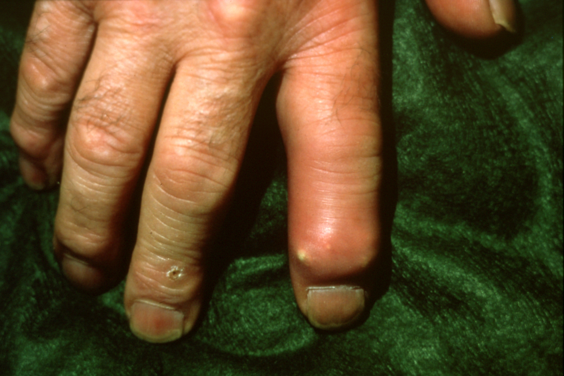
  • Gout at DIP presenting like a paronychia of the index finger.
    Gout at DIP presenting like a paronychia of the index finger.
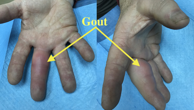
  • Acute Gout Long Finger causing pain and decreased range of motion
    Acute Gout Long Finger causing pain and decreased range of motion
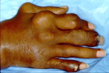
  • Severe uncontrolled chronic tophaceous gout
    Severe uncontrolled chronic tophaceous gout
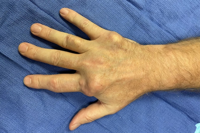
  • Tophaceous gout in right hand of a 57 year old male beer drinker.
    Tophaceous gout in right hand of a 57 year old male beer drinker.
Pathoanatomy Photos and Related Diagrams
Pathoanatomy Images
  • Surgically excised tophus
    Surgically excised tophus
Symptoms
Pain and swelling
Joint Tenderness
Erythema with warm skin
Decreased range of motion in affected joint(s)
Typical History

The typical patient is a 47-year-old obese man with hypertension and diabetes. He experienced his first flare of gout in his mid-30s, which was defined by pain, swelling, redness, and tenderness in his big toes and knees. Although he saw a doctor after having several more flares during that time, he did not make any significant modifications to his lifestyle, and the flares continued. More recently, the gout began to affect several DIP joints in his right hand, and symptoms prevented him from functioning normally, which led him to seek out medical advice.

Positive Tests, Exams or Signs
Work-up Options
Images (X-Ray, MRI, etc.)
Gout X-rays
  • AP X-ray of moderate gout of DIP joint. Note erosions (asymmetric punched-out defects)
    AP X-ray of moderate gout of DIP joint. Note erosions (asymmetric punched-out defects)
  • Lateral X-ray of moderate gout of DIP joint. Note erosions (asymmetric punched-out defects)
    Lateral X-ray of moderate gout of DIP joint. Note erosions (asymmetric punched-out defects)
  • Severe tophaceous gout with joint destruction and bone loss
    Severe tophaceous gout with joint destruction and bone loss
  • Severe tophaceous gout with DIP joint destruction and bone loss (arrow)
    Severe tophaceous gout with DIP joint destruction and bone loss (arrow)
  • Severe tophaceous gout with DIP joint destruction and phalangeal bone loss (arrow)
    Severe tophaceous gout with DIP joint destruction and phalangeal bone loss (arrow)
Treatment Options
Conservative
  • Gout management should be multidisciplinary and involve treatment of acute attacks, reduction of excess uric acid, and prophylactic therapy to prevent flares.2. The primary strategy for long-term management of gout is to reduce the serum urate to a concentration that achieves dissolution of MSU crystals. Therapy should be started as early as possible, and patients should have an action plan and supply of drugs to facilitate this approach.1
  • Nonsteroidal anti-inflammatory drugs (NSAIDs) are the first line of treatment for acute gout flares. NSAID's may be contraindicated in patients with kidney impairment, cardiovascular disease, or a history of gastrointestinal disease. Indomethacinis a commonly used NSAID but others are equally as affective.1
  • Colchicine
    • Colchicine is an alternative to NSAIDs for acute flares in patients with coexistent cardiovascular disease or a history of gastrointestinal bleeding.3
    • Colchicine also used to prevent recurrence in chronic gout. 1
  • Corticosteroids
    • Corticosteroids are indicated for patients who are unable to take NSAIDs or colchicine.
  • NSAIDs, colchicine, and corticosteroids can be used alone or in combination for more severe flares.1
  • Urate-lowering therapy
    • Recommended in the chronic stage, when a patient has more than two gouty episodes per year
    • Drug therapy must be individualized since treatment is lifelong and drug toxicities can occur2
    • Xanthine oxidase inhibitors inhibit urate production
      • Commonly given as first-line therapy for chronic gout attacks
      • Allopurinol is the most frequently used drug in this class
        • Must be used with caution in patients with renal impairment
        • Febuxostat
    • Uricosurics
      • Normalize renal urate excretion
      • Second-line option for patients who do not reach target serum urate concentrations with a xanthine oxidase inhibitor
      • Probenecid is a first-line uricosuric
        • Can be used as monotherapy or in combination with a xanthine oxidase inhibitor1
        • Benzbromarone
        • Lesinurad is a new RUAT1 inhibitor
    • Recombinant uricases catalyze the conversion of urate to allantoin
      • Pegloticase (IV every 2 wk) is typically reserved for patients with severe, refractory gout in whom target serum urate concentrations are not achieved or who cannot tolerate oral urate-lowering therapy
      • Rasburicase1
  • Urate-lowering lifestyle changes
    • Weight loss and diet modification, primarily increased vegetable and milk consumption and reduced consumption of meat, seafood, purine-rich foods of animal origin (eg, organ meats), and alcohol, especially beer
    • Indicated for patients with recurrent gout flares (>1 flare/y), tophi, stage 2 or worse chronic kidney disease, or kidney stones
    • Not recommended for people with asymptomatic hyperuricemia
    • Important to designate a target serum urate concentration based on disease severity1,2
  • Rest, topical ice, and elevation recommended to reduce symptoms during acute attacks
  • Elasticated wrapping and splinting aids may be helpful for stabilizing soft tissues, obliterating dead space, and stabilizing unstable distal joints5
Operative
  • May be necessary if functional disability persists despite aggressive medical management
  • Surgery should be focused on restoring joint and tendon mobility, nerve decompression, preventing skin breakdown, improving cosmesis, and reducing pain2
  • Carpal tunnel release
    • Combined with tendon debridement and used for addressing acute carpal tunnel syndrome during acute gouty attacks
  • Arthrodesis
    • Considered a safe and effective treatment for patients with severe involvement of the PIP and DIP joints.
    • Wrist arthrodesis may be needed for severe articular destruction and combined with excision of the lower end of the ulna2
  • Extensor tendon reconstruction is reserved reserved for patients with large tophi who may have involvement of other joints in the same digit or hand.
  • Other Surgical Options include:
    • Surgical irrigation is used to wash out MSU crystals
    • Debridement of septic joints
    • Debulking of painful or disfiguring tophi
    • Tendon transfer
    • Local synovectomy
    • Tophectomy
    • Amputation should only be considered in rare instances in which function is significantly impaired by pain, uncontrolled drainage, and infection, or if digital enlargement compromises adjacent digital function2
    • After surgery, early rehabilitation of early tendon motion in the fingers is critical to optimize final outcomes
Treatment Photos and Diagrams
Gout Surgical Treatment
  • Gouty painful DIP treat with arthrodesis
    Gouty painful DIP treat with arthrodesis
Complications
  • Infection
  • Delayed healing
  • Wound hematoma
  • Tendon ruptures
  • Joint destruction
Outcomes
  • The use of 0.6 mg prophylactic colchicine 1-2 times daily for the first six months of therapy has been found to decrease the likelihood and severity of flares2
  • Evidence suggests that NSAIDs are effective within the first several days of an acute gout flare, but no clinically significant differences in outcome or adverse effects have been reported among different NSAIDs3
  • Intra-articular corticosteroid injections seem to be effective for single joint involvement when there is no clinical concern for infection3
  • Pegloticase has been found to result in a profound reduction in serum urate, with rapid improvements in musculoskeletal function, health-related quality of life, pain, and tophus burden1
Key Educational Points
  • Ultrasound offers good diagnostic accuracy and should play a role in diagnosing gout and differentiating it from other arthritic diseases7
  • The prevalence and annual incidence of gout has been increasing for several reasons, include large-scale changes in diet, lifestyle and use of alcohol and diuretics1
  • Although the central cause of gout is well known and effective treatments are available, many uncertainties remain; for example:

    It’s not clear why some individuals with hyperuricemia develop MSU crystal deposition and others do not.
    Why MSU crystals deposit preferentially at specific sites and why deposited crystals can be present in the joint without clinically apparent inflammation.
    Causal relations between hyperuricemia and comorbid disorders such as hypertension, cardiovascular disease, and other features of metabolic syndrome are debated.
    At present, urate-lowering therapy is primarily recommended for patients with frequent flares or tophi, but the benefits of earlier initiation of urate-lowering therapy are unknown and will require careful analysis.
    The most important question about gout management may be how to improve long-term use of effective urate-lowering therapy.1
     
  • If left untreated, an acute gout flare resolves over 3-10 days, followed by an asymptomatic phase known as the intercritical period; as the disease progresses, these pain-free intervals become shorter and episodes of acute flares become more frequent6
  • The incidence of symptomatic tophaceous gout affecting the hand and wrist appears to be declining with improved medical management; however, about 5% of patients are recalcitrant to medical therapy and eventually need surgery2
  • Elective surgery should be done through a stable soft-tissue envelope, and involved areas that are clean and have intact skin coverage generally heal well with primary closure following debridement2
  • It is ill-advised to attempt excision of all tophaceous material when doing so would compromise structures important to hand function; tophaceous material that is firmly adherent to skin, tendon, and bone should be gently curetted or excised sharply, without compromising the integrity of the involved structure2
References

Cited

  1. Dalbeth. Gout. Lancet 2016;388(10055):2039-2052. PMID: 27112094
  2. Fitzgerald BT, Setty A, Mudgal CS. Gout affecting the hand and wrist. J Am Acad Orthop Surg 2007;15(10):625-35. PMID: 17916786
  3. Chimenti PC, Hammert WC. Medical management of acute gout. J Hand Surg Am 2012;37(10):2160-4. PMID: 22749478
  4. Wilczynski MC, Gelberman RH, Adams A, Goldfarb CA. Arthroscopic findings in gout of the wrist. J Hand Surg Am 2009;34(2):244-50. PMID: 19135809
  5. Mudgal CS. Management of tophaceous gout of the distal interphalangeal joint. J Hand Surg Br 2006;31(1):101-3. PMID: 16243417
  6. Zhu Y, Pandya BJ, Choi HK. Prevalence of gout and hyperuricemia in the US general population: the National Health and Nutrition Examination Survey 2007-2008. Arthritis Rheum 2011;63(10):3136-41. PMID: 21800283
  7. Lee YH, Song GG. Diagnostic accuracy of ultrasound in patients with gout: A meta-analysis. Semin Arthritis Rheum 2017 Epub. PMID: 29054295

New Articles

  1. Abhishek A, Roddy E, Doherty M. Gout - a guide for the general and acute physicians. Clin Med (Lond) 2017;17(1):54-59. PMID: 28148582
  2. So AK. Why better treatment of gout is needed. Clin Exp Rheumatol 2016;34(4 Suppl 98):63-5. PMID: 27586807

Reviews

  1. Ragab G, Elshahaly M, Bardin T. Gout: An old disease in new perspective - A review. J Adv Res 2017;8(5):495-511. PMID: 28748116
  2. Wilson L, Saseen JJ. Gouty Arthritis: A Review of Acute Management and Prevention. Pharmacotherapy 2016;36(8):906-22. PMID: 27318031

Classics

  1. Balfour W. Cases of Gout, with Observations. Med Chir J Rev 1818;5(27):191-196. PMID: 29257598
  2. Budd W. Researches on Gout. Med Chir Trans 1855;38:233-246.3. PMID: 20896045