Hand Surgery Source

THUMB PROXIMAL PHALANX FRACTURE

Introduction

Fracture Nomenclature for Thumb Proximal Phalanx Fracture

Hand Surgery Resource’s Diagnostic Guides describe fractures by the anatomical name of the fractured bone and then characterize the fracture by the Acronym:

In addition, anatomically named fractures are often also identified by specific eponyms or other special features.

For the Thumb Proximal Phalanx Fracture, the historical and specifically named fractures include:

Ulnar collateral ligament avulsion fracture/bony skier’s thumb/Gamekeeper's fracture

RCL avulsion fracture

MP joint fracture-dislocation with volar plate avulsion fracture

MP joint sesamoid bone fracture

By selecting the name (diagnosis), you will be linked to the introduction section of this Diagnostic Guide dedicated to the selected fracture eponym.


 Fractures of the proximal phalanx of the thumb are less common than those of the thumb distal phalanx and metacarpal. These injuries are particularly prevalent in sports and are often the result of direct blunt trauma or from a fall on an outstretched hand (FOOSH).1

Definitions

  • A thumb proximal phalanx fracture is a disruption of the mechanical integrity of the proximal phalanx.
  • A thumb proximal phalanx fracture produces a discontinuity in the proximal phalanx contours that can be complete or incomplete.
  • A thumb proximal phalanx fracture is caused by a direct force that exceeds the breaking point of the bone.

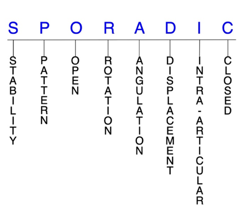
Hand Surgery Resource’s Fracture Description and Characterization Acronym

SPORADIC

S – Stability; P – Pattern; O – Open; R – Rotation; A – Angulation; D – Displacement; I – Intra-articular; C – Closed


S - Stability (stable or unstable)

  • Universally accepted definitions of clinical fracture stability is not well defined in the hand surgery literature.2-4
  • Stable: fracture fragment pattern is generally nondisplaced or minimally displaced. It does not require reduction, and the fracture fragments’ alignment is maintained with simple splinting. However, most definitions define a stable fracture as one that will maintain anatomical alignment after a simple closed reduction and splinting. Some authors add that stable fractures remain aligned, even when adjacent joints are put to a partial range of motion (ROM).
  • Unstable:  fracture will not remain anatomically or nearly anatomically aligned after a successful closed reduction and simple splinting. Typically unstable thumb proximal phalanx fractures have significant deformity with comminution, displacement, angulation, and/or shortening.

P - Pattern

  • Thumb proximal phalanx head: oblique, transverse, or comminuted; can involve the interphalangeal (IP) joint; these are intra-articular fractures that affect one or both condyles of the thumb proximal phalanx head, with or without displacement; displaced fractures can affect joint congruity.
  • Thumb proximal phalanx shaft: transverse, oblique, or comminuted with or without shortening.
  • Thumb proximal phalanx base: can involve the metacarpophalangeal (MP) joint; fractures at the thumb proximal phalanx base are frequently concomitant with avulsions fractures of ligaments associated with the MP joint.5-7

O - Open

  • Open: a wound connects the external environment to the fracture site. The wound provides a pathway for bacteria to reach and infect the fracture site. As a result, there is always a risk in developing osteomyelitis. Therefore, open fractures of the thumb proximal phalanx require antibiotics with surgical irrigation and wound debridement.2,8,9

R - Rotation

  • Thumb proximal phalanx fracture deformity can be caused by proximal rotation of the fracture fragment in relation to the distal fracture fragment.
  • Degree of malrotation of the fracture fragments can be used to describe the fracture deformity; this is not a common type of fracture deformity in the thumb proximal phalanx.

A - Angulation (fracture fragments in relationship to one another)

  • Angulation is measured in degrees after identifying the direction of the apex of the angulation.
  • Straight: no angulatory deformity
  • Angulated: bent at the fracture site
  • Example: unlike the finger phalanges, some angular displacement or malunion is acceptable in thumb proximal phalanx fractures due to the compensatory motion of the MP joint; therefore, angular deformities of up to 20° in the frontal plane and 30° in the lateral plane may be functionally well tolerated in these fractures.5

D - Displacement (Contour)

  • Displaced: disrupted cortical contours (eg, thumb proximal phalanx shaft fractures can be displaced or translated partially or completely)
  • Nondisplaced: fracture line defining one or several fracture fragments; however, the external cortical contours are not significantly disrupted

I - Intra-articular involvement

  • Fractures that enter a joint with one or more of their fracture lines. Intra-articular fractures of the thumb proximal phalanx require special attention due to the digit’s indispensible role in hand function.9
  • Thumb proximal phalanx fractures can have fragment involvement with the IP or MP joints.
  • If a fracture line enters a joint but does not displace the articular surface of the joint, then it is unlikely that this fracture will predispose to posttraumatic osteoarthritis. If the articular surface is separated or there is a step-off in the articular surface then the congruity of the joint will be compromised and the risk of posttraumatic osteoarthritis increases significantly.

C - Closed

  • Closed: no associated wounds; the external environment has no connection to the fracture site or any of the fracture fragments.2-4

Thumb distal phalanx fractures: named fractures, fractures with eponyms and other special fractures

Ulnar collateral ligament avulsion fracture/bony skier’s thumb/Gamekeeper's fracture

  • Injuries to the ulnar collateral ligament (UCL) occur due to hyperabduction and forceful radial deviation of the thumb MP joint, and they are particularly common in contact sports and skiing.8
  • An acute injury to the UCL is commonly called “skier’s thumb,” since it often results from a ski pole keeping the thumb in an abducted position, which increases its risk for excessive radial deviation. Skier’s thumb can also occur in ball or stick sports or from a FOOSH.
    • The estimated annual incidence of skier’s thumb in the US is ~200,000.10
    • The majority of skier’s thumb injuries result in a distal avulsion fracture of the UCL, and about 50% of all UCL injuries will have an associated fracture at the base of the thumb proximal phalanx. The term “bony skier’s thumb” may be used to describe a UCL avulsion fracture.6
  • A chronic UCL injury is commonly referred to as “gamekeeper’s thumb,” as the injury was first identified in Scottish gamekeepers who experienced chronic ligament insufficiency consequential of their method for breaking the necks of rabbits between the thumb and index finger.
    • This chronic injury can also result in a UCL avulsion fracture, and is approached in a similar manner to its conservative counterpart.6
  • UCL injuries occur significantly more frequently than radial collateral ligament (RCL) injuries.5
  • Displacement of the UCL proximal and superficial to the leading edge of the adductor pollicis aponeurosis is termed a Stener lesion. In these injuries, the aponeurosis of the adductor pollicis is interposed between the UCL and its insertion on the proximal phalanx, which prohibits healing.
    • Accurate diagnosis of Stener lesions is often a diagnostic dilemma, since they often occur in the absence of a substantial bony fragment.5
    • Some investigators have described a palpable mass proximal to the MP joint as a sign of a Stener lesion and occasionally an associated avulsion fracture with the fragment proximal to the location of the adductor hood will indicate a “bony Stener” lesion.6

Imaging

  • Plain anteroposterior and lateral X-ray views of the thumb are recommended.
  • Arthrogram
  • Ultrasound
  • MRI

Treatment

  • Treatment for thumb UCL avulsion fractures is controversial, since many of these injuries are capable of primary healing if minimally displaced, and the available literature supports both conservative and surgical management strategies.5
  • In general, the research suggests that undisplaced or minimally displaced avulsion fractures with no joint instability should be managed conservatively.11
    • Conservative treatment should involve a thumb spica cast that includes the wrist until initial inflammation has resolved, followed by a thermoplastic splint that allows movement at the IP joint.
    • The position of the splint involves holding the MP joint in slight flexion with gentle stressing in ulnar deviation. During immobilization, the patient should undergo supervised hand therapy.12
    • Gentle flexion/extension exercises can begin after 4 weeks with the splint being worn between sessions, and the splint should remain on at all times apart from during therapy sessions for 6 weeks. Progressive strengthening exercises may begin after 8 weeks, and unrestricted activity is allowed after 12 weeks.12
  • On the other hand, ORIF should be performed if: 1) 20% or greater of the joint surface is involved, 2) there is considerable fracture displacement, or 3) there is substantial instability with UCL testing. The goal of surgery is stable fixation that permits early motion.5,11
    • The presence of a Stener lesion is another indication for surgical intervention.5
    • If the fracture fragment is displaced more than 2 mm and the MP joint is unstable to stress, stability needs to be restored surgically.
  • If the fracture fragment is small or breaks during internal fixation, it can be removed and the ligament reinserted with a pull-out suture or suture anchor.
  • Larger fragments can be fixed with either K-wires or a small screw. The repair is protected with a transarticular smooth K-wire and thumb spica cast immobilization for 4-6 weeks.1
  • Tension band wiring is a useful technique for fragments not large enough to support a screw, while suture anchors are recommended for smaller and comminuted fractures.11
  • If there is any doubt about joint stability, stress testing should be performed under a median and radial nerve wrist block with the thumb at 30° MP flexion. If the joint opens 35° or more with radial deviation stress, then surgery is indicated.11
  • For some surgeons, the presence of a displaced avulsion fracture is an indication for surgical intervention even if it is not displaced enough to be superficial to the adductor aponeurosis.13

Complications

  • MP joint instability
  • Nonunion
  • Impaired grip/pinch strength
  • Posttraumatic osteoarthritis

Outcomes

  • Several studies have reported complete stability and no pain in a 63-100% of patients after being treated conservatively for nondisplaced UCL avulsion fractures.13
  • Good outcomes were found in one study of 30 patients with UCL avulsion fractures treating conservatively, with 19 patients pain-free, 10 with mild pain, and 1 with moderate pain. Impairments in stability and grip/pinch strength were also rare. The authors of this study therefore recommend conservative treatment for most acute UCL avulsion fractures.14
    • Conversely, another study found identified potential problems of conservatively treating small, minimally displaced UCL avulsion fractures, with 9 patients initially treated with cast immobilization experiencing persistent pain and diminished pinch and grip strength after at least 6 weeks in the cast. Late UCL reconstruction was subsequently performed in all patients with K-wire fixation and pull-through sutures, and subsequent outcomes were good.15
  • Another study found that a hook plate construct was biomechanically superior and provided superior fixation of UCL avulsion fractures compared to the suture anchor construct. Hook plating may therefore allow an earlier return to forceful pinch activities and also provide a greater resistance to catastrophic failure in the early postoperative period for non-compliant or highly active patients.7

RCL avulsion fracture

  • The RCL plays a significant role in stabilizing the MP joint, but injuries and avulsion fractures of this ligament are far less common than those of the UCL. RCL avulsion fractures typically occur due to forced adduction or torsion of the flexed thumb, which can occur in sports like handball or soccer, or from a FOOSH. Evaluation, diagnosis, and management is similar to that for UCL injuries, but there are several key differences between the two:16
    • In contrast with the adductor aponeurosis, the abductor aponeurosis on the radial side of the thumb is broad and does not become interposed between the ligament and avulsed insertion site. As a result, equivalents of Stener lesions in RCL avulsion fractures are extremely rare.17
    • Similarly, the location of RCL injuries is more variable. Most frequently, the ligament is avulsed from its proximal origin on the metacarpal as opposed to its distal insertion on the proximal phalanx.
    • Midsubstance tears of the RCL are more common than midsubstance tears of the UCL.17
    • Joint subluxation is more common with RCL injuries owing to the adductor pollicis insertion on the proximal phalanx and ulnar sesamoid. These insertions lie volar to the MP joint axis of rotation, causing a volar and ulnar-deforming force on the proximal phalanx.6

Imaging

  • Anteroposterior and lateral X-ray views are recommended.
  • Arthrogram
  • Ultrasound
  • MRI

Treatment

  • Both conservative and surgical intervention strategies have been utilized for RCL avulsion fractures, with both options being well supported in the literature if the fracture is minimally displaced.6
    • Non-displaced, non-rotated avulsed fragments are usually treated non-surgically, while displaced or rotated fragments should undergo surgical treatment. However, consistent recommendations and algorithms for optimal management of these injuries are yet to be delineated.6
    • Though rare, Stener-like lesions on the radial side are also indications for surgery.6
    • To date, two methods for surgical repair are mainly conducted: reduction and fixation or excision of the bony fragment, depending on fragment characteristics.16
    • Rigid fixation of the fragment can be performed with a tension band construct or a 1.5-mm interfragmentary screw. If there are multiple small fragments, excision of those fragments with reattachment of the insertion of the RCL can be performed.6
    • Osteosynthesis with ORIF is indicated except when a fragment is not displaced and the size of a fragment is very small.18

Complications

  • MP joint instability
  • Impaired pinch/grip strength
  • Posttraumatic osteoarthritis

Outcomes

  • Osteosynthesis was found to lead to bony union and good functional results with no complications and high satisfaction rates in patients with both UCL and RCL avulsion fractures.18
  • In one study on patients with RCL avulsion fractures, 9 were treated surgically and 21 were treated non-surgically following a devised treatment algorithm. Outcomes were similar between groups, with 90.5% of patients in both groups being graded as “excellent” after treatment. There were also no significant between-group differences in post-treatment ROM.16

MP joint fracture-dislocation with volar plate avulsion fracture

  • Traumatic dislocation of the thumb MP joint is considered a rare injury, but this may be due to it being underreported in the literature. The usual mechanism of injury is a FOOSH that causes forcible hyperextension of the joint. Dorsal dislocations are more frequent than volar dislocations, and MP joint dislocations are less common than IP dislocations.
    • Concomitant fractures of the base of the proximal phalanx and metacarpal head occur in about 50% of MP joint dislocations.
    • A fracture-dislocation of the thumb MP can also lead to avulsion of the volar plate, which is avulsed from its attachment on the metacarpal neck during MP hyperextension.
  • In a simple dislocation, the volar plate is not interposed within the MP joint, and the base of the proximal phalanx remains in contact with the articular surface of the metacarpal head.
  • In a complex dislocation, the volar plate becomes inserted into the MP joint, and open reduction is usually needed.19

Imaging

  • Anteroposterior, lateral, and oblique X-ray views are recommended.

Treatment

  • Unlike UCL avulsion fractures, a volar plate avulsion is considered a stable injury, and these injuries should be treated conservatively whenever feasible.11
  • Some simple fracture-dislocations can be reduced non-surgically with initial MP hyperextension followed by dorsal pressure on the proximal phalanx in conjunction with wrist flexion.19
  • Complex MP fracture-dislocations typically require open surgical reduction.20
    • Simple distraction as a reduction maneuver is usually unsuccessful and can inadvertently convert a reducible dislocation into an irreducible one, as traction on the MP joint can draw the entire volar plate dorsally so that it can be completely folded between the base of the thumb proximal phalanx and metacarpal head.19
  • When the volar plate becomes entrapped within the MP joint in an avulsion fracture, a closed reduction maneuver cannot be completed.19
  • Because the anatomy of the thumb is different from that of the other fingers, dislocations requiring surgical intervention frequently involve interposition of the volar plate, sesamoid bones, or flexor pollicis longus tendon.19

Complications

  • Posttraumatic osteoarthritis
  • Osteonecrosis
  • MP joint arthofibrosis/stiffness
  • Decreased ROM

Outcomes

  • Dorsal dislocations of the thumb MP joint are treated by the same reduction maneuver as the other fingers, but treatment has been found to be more likely to be successful in the thumb.19

MP joint sesamoid bone fracture

  • Two sesamoid bones are present at the MP joint of thumb in more than 99% of people, and their function is probably to protect and stabilize the flexor tendons and intrinsic muscles of the hand. Fractures of either of these bones are very uncommon and can be easily missed in a diagnosis.21
  • The most common mechanism of injury for MP joint sesamoid bone fractures is a violent hyperextension or hyperabduction of the thumb after a FOOSH, which may be related to sports. They may also result from direct trauma.21,22
  • The volar plate can also be torn with or without an avulsion fracture in these injuries.22
  • Sesamoid bone fractures are generally classified into two types:
  • Type 1: the volar plate and MP joint remain intact.
  • Type 2: rupture of the volar plate with associated thumb hyperextension.21

Imaging

  • Since sesamoid fractures may not be evident on routine anteroposterior and lateral views, oblique views should be obtained when this type of fracture is suspected.
  • Ultrasound may be particularly useful to detect sesamoid fractures that are clinically or radiographically overlooked.
  • CT scan may also be necessary.

Treatment

  • It is important to diagnose and treat sesamoid bone fractures as early as possible, since these injuries can be easily missed.21
  • Type 1 sesamoid bone fractures can be treated conservatively with immobilization of the thumb and the MP joint in 30° of flexion for 2-4 weeks. Taping may also be sufficient. If pain and discomfort persist after adequate conservative treatment, excision of the bone may be needed.21,23
  • Type 2 fractures may require surgical reduction of the MP joint and sesamoid fracture and repair of the volar plate if the fracture is complicated and instability of the MP joint is clinically demonstrated.21,23
  • Other recommendations call for conservative treatment of closed sesamoid fractures and surgical treatment for open fractures, in which the fracture fragments are approximated to reinforce the volar plate repair to prevent a hyperextension deformity of the MP joint.23

Complications

  • Stiffness of MP joint

Outcomes

  • The prognosis for a sesamoid bone fracture is good when it is properly diagnosed at an early stage, but poor functional outcomes are common when the injury is missed.
  • Taping has been associated with positive overall outcomes and no complications in one study.23

Related Anatomy

  • The thumb proximal phalanx consists of a distal phalangeal head that articulates at the IP joint with the distal phalanx, a narrow diaphyseal shaft, a proximal metaphysis, and a base that articulates at the MP joint with the metacarpal. The base of the thumb proximal phalanx also has a dorsal and volar lip.
  • The ligaments associated with the thumb proximal phalanx at the IP and MP joints are the joint capsule, the UCL and RCL (proper and accessory collaterals), and the volar plates.
  • Tendon attachments include the abductor pollicis brevis, flexor pollicis brevis, and opponens pollicis.

Incidence and Related injuries/conditions

  • Metacarpal and phalangeal fractures account for nearly half of all hand injuries that present to the emergency room.24
  • Phalangeal fractures account for 23% of all below-elbow fractures.25
  • Fractures of the thumb are most common in children and the elderly, with the thumb being the most commonly fractured tubular bone in elderly patients.26
    • Thumb proximal phalanx fractures most commonly occur secondary to sports in younger patients, secondary to labor in middle-aged patients, and secondary to falls and motor vehicle accidents in older individuals.9
  • The estimated annual incidence of skier’s thumb in the U.S. is ~200,000.10
ICD-10 Codes

GAMEKEEPER'S THUMB FRACTURE

Diagnostic Guide Name

GAMEKEEPER'S THUMB FRACTURE

ICD 10 Diagnosis, Single Code, Left Code, Right Code and Bilateral Code

DIAGNOSIS SINGLE CODE ONLY LEFT RIGHT BILATERAL (If Available)
FRACTURE THUMB - PROXIMAL PHALANX (GAMEKEEPER'S THUMB FRACTURE)        
- DISPLACED   S62.512_ S62.511_  
- NONDISPLACED   S62.515_ S62.514_  

Instructions (ICD 10 CM 2020, U.S. Version)

THE APPROPRIATE SEVENTH CHARACTER IS TO BE ADDED TO EACH CODE FROM CATEGORY S62
  Closed Fractures Open Type I or II or Other Open Type IIIA, IIIB, or IIIC
Initial Encounter A B C
Subsequent Routine Healing D E F
Subsequent Delayed Healing G H J
Subsequent Nonunion K M N
Subsequent Malunion P Q R
Sequela S S S

ICD-10 Reference

Reproduced from the International statistical classification of diseases and related health problems, 10th revision, Fifth edition, 2016. Geneva, World Health Organization, 2016 https://apps.who.int/iris/handle/10665/246208

THUMB PROXIMAL PHALANX FRACTURE

Diagnostic Guide Name

THUMB PROXIMAL PHALANX FRACTURE

ICD 10 Diagnosis, Single Code, Left Code, Right Code and Bilateral Code

DIAGNOSIS SINGLE CODE ONLY LEFT RIGHT BILATERAL (If Available)
FRACTURE THUMB - PROXIMAL PHALANX (GAMEKEEPER'S THUMB FRACTURE)        
- DISPLACED   S62.512_ S62.511_  
- NONDISPLACED   S62.515_ S62.514_  

Instructions (ICD 10 CM 2020, U.S. Version)

THE APPROPRIATE SEVENTH CHARACTER IS TO BE ADDED TO EACH CODE FROM CATEGORY S62
  Closed Fractures Open Type I or II or Other Open Type IIIA, IIIB, or IIIC
Initial Encounter A B C
Subsequent Routine Healing D E F
Subsequent Delayed Healing G H J
Subsequent Nonunion K M N
Subsequent Malunion P Q R
Sequela S S S

ICD-10 Reference

Reproduced from the International statistical classification of diseases and related health problems, 10th revision, Fifth edition, 2016. Geneva, World Health Organization, 2016 https://apps.who.int/iris/handle/10665/246208

UCL AVULSION FRACTURE (SKIER'S THUMB)

Diagnostic Guide Name

UCL AVULSION FRACTURE (SKIER'S THUMB)

ICD 10 Diagnosis, Single Code, Left Code, Right Code and Bilateral Code

DIAGNOSIS SINGLE CODE ONLY LEFT RIGHT BILATERAL (If Available)
UCL AVULSION FRACTURE (SKIER'S THUMB)   S63.642_ S63.641_  

Instructions (ICD 10 CM 2020, U.S. Version)

THE APPROPRIATE SEVENTH CHARACTER IS TO BE ADDED TO EACH CODE FROM CATEGORY S68
A - Initial Encounter
D - Subsequent Routine Healing
S - Sequela

ICD-10 Reference

Reproduced from the International statistical classification of diseases and related health problems, 10th revision, Fifth edition, 2016. Geneva, World Health Organization, 2016 https://apps.who.int/iris/handle/10665/246208

Clinical Presentation Photos and Related Diagrams
  • Bicondylar non-displaced intra-articular thumb proximal phalanx fracture
    Bicondylar non-displaced intra-articular thumb proximal phalanx fracture
  • Condylar displaced intra-articular thumb proximal phalanx fracture
    Condylar displaced intra-articular thumb proximal phalanx fracture
  • Thumb angulated proximal phalanx shaft fracture
    Thumb angulated proximal phalanx shaft fracture
  • Non-displaced intra-articular thumb proximal phalanx base fracture
    Non-displaced intra-articular thumb proximal phalanx base fracture
  • Displaced intra-articular thumb proximal phalanx base fracture at the radial collateral insertion. Note the articular surface on the fracture fragment is rotated out of the joint. This will require CRIF or ORIF.
    Displaced intra-articular thumb proximal phalanx base fracture at the radial collateral insertion. Note the articular surface on the fracture fragment is rotated out of the joint. This will require CRIF or ORIF.
  • Displaced intra-articular thumb proximal phalanx base fracture (Gamekeeper's Fracture) at the ulnar collateral insertion. Note the articular surface on the fracture fragment is rotated out of the joint. This will require CRIF or ORIF.
    Displaced intra-articular thumb proximal phalanx base fracture (Gamekeeper's Fracture) at the ulnar collateral insertion. Note the articular surface on the fracture fragment is rotated out of the joint. This will require CRIF or ORIF.
Symptoms
Pain in the injured thumb
Swelling in the injured thumb
Ecchymosis in the injured thumb
Deformity in the injured thumb
Loss of range of motion
Typical History

A classic patient with a thumb proximal phalanx fracture is a 35-year-old, right-handed male who was skiing at a fast pace when he planted his left pole in a firm section of snow and caught an edge of his right ski. The man’s body continued moving forward and he fell to the ground while the pole remained planted in the snow, and as a result, the strap of his pole caught his left thumb and forced it into hyperabduction and excessive radial deviation. The incident resulted in a UCL avulsion fracture at the base of the proximal phalanx, which led to immediate pain and swelling of the injured area.

Positive Tests, Exams or Signs
Work-up Options
Images (X-Ray, MRI, etc.)
Thumb Proximal Phalanx X-rays
  • Thumb proximal phalanx angulated base fracture AP and oblique views
    Thumb proximal phalanx angulated base fracture AP and oblique views
  • Thumb proximal phalanx angulated base fracture lateral view. Fracture will require closed reduction and cast.
    Thumb proximal phalanx angulated base fracture lateral view. Fracture will require closed reduction and cast.
  • Thumb intraarticular radial collateral avulsion fracture with malrotation
    Thumb intraarticular radial collateral avulsion fracture with malrotation
  • Thumb intraarticular radial collateral avulsion fracture with cartilage surface rotated into the fracture site (arrow).
    Thumb intraarticular radial collateral avulsion fracture with cartilage surface rotated into the fracture site (arrow).
  • Thumb intraarticular ulnar collateral avulsion fracture, Gamekeeper's FX, site (arrow).
    Thumb intraarticular ulnar collateral avulsion fracture, Gamekeeper's FX, site (arrow).
Treatment Options
Treatment Goals
  • When treating closed thumb proximal phalanx fractures, the treating surgeon has 4 basic goals:2,9
    1. A thumb with a normal appearance. The X-ray may not need to be perfect but the thumb should have no obvious deformity (ie, the thumb looks normal!)
    2. Avoid thumb stiffness by maintaining a normal functional ROM (ie, the thumb works!)
    3. The thumb is not painful (ie, the thumb does not hurt!)
    4. Congruent joint surface with none-to-minimal joint surface irregularities (ie, the joint does not develop early posttraumatic arthritis!)
    5. Fracture care should minimize the risk for infection and osteomyelitis (This is an additional goal outside of the four basic goals, but it is mandatory for open fractures)
  • One additional goal is mandatory for open fractures:
  • Many thumb fractures are treated similarly to finger fractures, but special consideration is needed since the thumb is distinct from the other digits: Whereas adjacent fingers can compensate for one another, there is no surrogate for the thumb. Therefore, intra-articular fractures of the thumb proximal phalanx demand special care.6,9
Conservative
  • Most thumb proximal phalanx fractures can be treated without surgical treatment.10
  • The typical closed, nondisplaced, minimally angulated, thumb proximal phalanx fracture without significant malrotation can be managed in an aluminum plaster or fiberglass or custom splint.
    • Safe splinting of the thumb holds all joints in extension and the thumb in abduction.9
  • Thumb proximal phalanx fractures usually do not require that the thumb be included in a short-arm cast.
  • Even thumb proximal phalanx fractures that require a reduction to correct fracture-related deformity usually can be held in anatomic or near-anatomic alignment with a splint without internal or external surgical fixation.
  • Other indications for determining whether conservative or surgical treatment is appropriate include the following:
    • Fractures involving <30% of the joint are typically stable and can be treated with extension splinting.
    • Fractures with persistent volar subluxation, joint incongruity, or >50% joint involvement should be addressed surgically.6
  • Managing extra-articular thumb phalangeal fractures differs from that of finger phalangeal fractures in that some angular displacement or malunion is acceptable due to the compensatory motion of the MP joint, and in the thumb proximal phalanx, angular deformities ≤20° in the sagittal plane and 30° in the coronal plane may be functionally well tolerated.5
  • Early rehabilitation and physical therapy to improve finger ROM and reduce stiffness should also be carried out in most cases regardless of the treatment used, but it is particularly necessary in oblique and complex proximal phalanx fractures managed conservatively.27,28
Operative
  • Surgical treatment of thumb proximal phalanx fractures must always be an individualized therapeutic decision. However, surgical thumb proximal phalanx fracture care is most frequently recommended when:
    1. Closed reduction fails or the simple splint or cast immobilization does not maintain the reduction. For these irreducible or unstable fractures, operative treatment is recommended to achieve the 4 treatment goals of fracture care.
    2. There is a significantly displaced base of thumb proximal phalanx fracture involving the MP joint, surgical fracture care may be required (eg, displaced avulsion fractures of the UCL, RCL, or volar plate).
    3. Open thumb proximal phalanx fractures require surgical care in the form of irrigation and debridement to prevent chronic infection or osteomyelitis.
  • Percutaneous fixation is usually appropriate for most thumb proximal phalanx fractures, and simple fractures rarely require ORIF.9
    • Fixation depends on the fracture type and surgeon preference, but K-wires or interfragmentary screw fixation is often adequate. Stable fixation offers the advantages of earlier mobilization.5
    • Thumb proximal phalanx nonunions may also be surgically managed with percutaneous compression screws.10
    • Displaced spiral or oblique fractures of the head and neck of the thumb proximal phalanx may be treated by percutaneous pinning or by open reduction with either K-wires or interfragmentary screws.1
    • If open reduction of a thumb proximal phalanx fracture is required, the fracture is exposed through a dorsal “Y”-shaped incision with the extensor pollicis longus insertion left intact.1

Post-treatment Management

  • The care and precautions related to immobilization devices for the thumb proximal phalanx fracture must be carefully reviewed with the patient. Patients should be educated regarding care and precautions. Patients should know that pain, especially increasing pain, numbness, tingling, skin irritation, splint loosening, or excessive splint tightness are red flags and should be reported to the surgeon or his team.
  • Pain should be managed with properly fitting splints, reassurance, elevation, ice in the initial post-fracture period, and mild pain medications. Patients should be encouraged to discontinue pain medication as soon as possible. Opioid use should be kept to a minimum.
  • Joints that are splinted for closed stable fractures are usually immobilized.
  • Fractures that require internal fixation can be mobilized after 4 weeks.
  • If an infection does occur, management should focus on eradicating sepsis with thorough debridement, appropriate antibiotics (eg, cephalosporin, penicillin), and fracture stabilization, followed by obtaining fracture union and regaining a functional extremity.1
  • Patients should be instructed to carefully exercise all joints in the injured hand that do not require immobilization. Patients usually can exercise on their own; however, signs of generalized thumb or hand stiffness are indications for referral to hand therapy (PT or OT).
Treatment Photos and Diagrams
Thumb Proximal Phalanx Fracture Treatment
  • Thumb intraarticular radial collateral avulsion fracture fixation AP view:  (1) Pin immobilizing MP joint; (2) Pins for internal fixation of fragment
    Thumb intraarticular radial collateral avulsion fracture fixation AP view: (1) Pin immobilizing MP joint; (2) Pins for internal fixation of fragment
  • Thumb intraarticular radial collateral avulsion fracture fixation Lateral view:  (1) Pin immobilizing MP joint; (2) Pins for internal fixation of fragment
    Thumb intraarticular radial collateral avulsion fracture fixation Lateral view: (1) Pin immobilizing MP joint; (2) Pins for internal fixation of fragment
CPT Codes for Treatment Options

Per an agreement between Hand Surgery Resource, LLC and the American Medical Association (AMA) users are required to accept the following End User Point and Click Agreement in order to view CPT content on this website.  Please read and then click "Accept" at the bottom to indicate your acceptance of the agreement.

End User Point and Click Agreement

CPT codes, descriptions and other data only are copyright 2019 American Medical Association (AMA). All rights reserved. CPT is a registered trademark of the AMA. The AMA reserves all rights to approve any license with any Federal agency.

You, as an individual, are authorized to use CPT only as contained in Hand Surgery Resource solely for your own personal information and only within the United States for non-commercial, educational use for the purpose of education relating to the fundamental principles of hand surgery and the common diseases, disorders and injuries affecting the human hand. You agree to take all necessary steps to ensure your compliance with the terms of this agreement.

Any use not authorized herein is prohibited, including by way of illustration and not by way of limitation, making copies of CPT for resale and/or license, transferring copies of CPT to any party not bound by this agreement, creating any modified or derivative work of CPT, or making any commercial use of CPT. License to use CPT for any use not authorized herein must be obtained through the AMA, CPT Intellectual Property Services, 330 N. Wabash Avenue, Chicago, IL 60611. Applications are available at the AMA Web site, http://www.ama-assn.org/go/cpt.

Common Procedure Name
Gamekeeper's repair
CPT Description
Primary repair collateral ligament metacarpophalangeal joint
CPT Code Number
26542
Common Procedure Name
ORIF proximal phalanx
CPT Description
Open treatment phalangeal shaft fracture proximal/mid finger/thumb w/wo fixation each
CPT Code Number
26735
CPT Code References

The American Medical Association (AMA) and Hand Surgery Resource, LLC have entered into a royalty free agreement which allows Hand Surgery Resource to provide our users with 75 commonly used hand surgery related CPT Codes for educational promises. For procedures associated with this Diagnostic Guide the CPT Codes are provided above. Reference materials for these codes is provided below. If the CPT Codes for the for the procedures associated with this Diagnostic Guide are not listed, then Hand Surgery Resource recommends using the references below to identify the proper CPT Codes.

 CPT QuickRef App.  For Apple devices: App Store. For Android devices: Google Play

 CPT 2021 Professional Edition: Spiralbound

Complications
  • Stiffness is the most common complication of hand fractures,2,9 but thumb proximal phalanx fractures can usually be immobilized rapidly enough to avoid this.
  • Malunion and fingertip deformity after thumb proximal phalanx fractures is rare but can occur, especially in open, severe, unstable fractures.29 Nonunions are also rare, and guidelines for treating these complications do not exist. Malunions with malrotation are also possible.30
  • Posttraumatic osteoarthritis can occur in the MP joint after some thumb proximal phalanx fractures (eg, UCL avulsion fracture).
  • Osteomyelitis of the thumb proximal phalanx is rare but can occur in open thumb proximal phalanx fractures, especially in patients with diabetes or in patients whose immune system is compromised.
  • Because of the compensatory movement of the adjacent joints, the thumb is more forgiving of residual deformity than the other digits. The thumb’s ROM also makes it additionally resistant to malunion.1,9
Outcomes
  • Most outcomes after thumb proximal phalanx fractures are very good.2,9 Fortunately, the complications noted above are very rare. Significant stiffness can usually be avoided because the IP joint of the thumb can be mobilized while the MP joint and proximal phalanx are splinted.
  • In general, the prognosis for thumb distal phalanx fractures and fracture-dislocations has a great deal to do with the amount of energy associated with the original injury.
    • High-energy injuries often produce comminution, articular surface damage, and extensive soft tissue injury, which predispose patients to degenerative changes and stiffness, and usually lead to poor outcomes.
    • Low-energy injuries with simple fracture patterns and limited soft tissue involvement are generally associated with much better prognosis.
Key Educational Points
  • Thumb proximal phalanx fractures must be mobilized before radiographic fracture healing is complete to avoid disabling stiffness.
  • Immobilization of thumb proximal phalanx fractures for >4 weeks is rarely needed.1
  • Today, most thumb proximal phalanx fractures can be treated without surgery.1,4
  • Underlying pathological conditions such as bone tumors like enchondromas and osteoporosis should be expected in fractures that occur from trivial trauma.
  • The functional needs of each patient must be considered when recommending treatment for finger fractures.
  • The management of extra-articular thumb phalanx fractures differs from that of finger phalanx fractures in that some angular displacement or malunion is acceptable due to the compensatory motion of the thumb MP joint.5
  • The thumb provides up to 40% of hand function, and total disability of the thumb can be devastating, as it equilibrates to a loss of 22% of bodily function.5
  • ROM at the thumb MP joint is the most variable in the human body and may even differ between the right and left hands in the same patient.5
References

New and Cited Articles

  1. Day CS. Fractures of the Metacarpals and Phalanges. In: Green DP, ed. Green's Operative Hand Surgery. Seventh ed. Philadelphia: Elsevier; 2016, pp. 231-77.
  2. Cheah AE, Yao J. Hand Fractures: Indications, the Tried and True and New Innovations. J Hand Surg Am 2016;41:712-22. PMID: 27113910
  3. Nesbitt KS, Failla JM, Les C. Assessment of instability factors in adult distal radius fractures. J Hand Surg Am 2004;29:1128-38. PMID: 15576227
  4. Walenkamp MM, Vos LM, Strackee SD, Goslings JC, Schep NW. The Unstable Distal Radius Fracture-How Do We Define It? A Systematic Review. J Wrist Surg 2015;4:307-16. PMID: 26649263
  5. Carlsen BT, Moran SL. Thumb trauma: Bennett fractures, Rolando fractures, and ulnar collateral ligament injuries. J Hand Surg Am 2009;34(5):945-52. PMID: 19411003
  6. Kadow TR, Fowler JR. Thumb Injuries in Athletes. Hand Clin 2017;33(1):161-173. PMID: 27886832
  7. Shin EH, Drake ML, Parks BG, Means KR Jr. Hook Plate Versus Suture Anchor Fixation for Thumb Ulnar Collateral Ligament Fracture-Avulsions: A Cadaver Study. J Hand Surg Am 2016;41(2):192-5. PMID: 26718070
  8. Ketonis C, Dwyer J, Ilyas AM. Timing of Debridement and Infection Rates in Open Fractures of the Hand: A Systematic Review. Hand (NY) 2017;12:119-26. PMID: 28344521
  9. Meals C, Meals R. Hand fractures: a review of current treatment strategies. J Hand Surg Am 2013;38:1021-31. PMID: 23618458
  10. Hinke DH, Erickson SJ, Chamoy L, Timins ME. Ulnar collateral ligament of the thumb: MR findings in cadavers, volunteers, and patients with ligamentous injury (gamekeeper's thumb). Am J Roentgenol 1994;163(6):1431-4. PMID: 7992741
  11. Husband JB, McPherson SA. Bony skier's thumb injuries. Clin Orthop Relat Res 1996;(327):79-84. PMID: 8641086
  12. Anderson D. Skier's thumb. Aust Fam Physician 2010;39(8):575-7. PMID: 20877752
  13. Stoop N, Teunis T, Ring D, Eberlin KR. Variation in the Rate of Surgery for Ulnar Collateral Ligament Injury of the Metacarpophalangeal Joint of the Thumb. Hand (NY) 2017;12(5):512-517. PMID: 28774189
  14. Kuz JE, Husband JB, Tokar N, McPherson SA. Outcome of avulsion fractures of the ulnar base of the proximal phalanx of the thumb treated nonsurgically. J Hand Surg Am 1999;24(2):275-82. PMID: 10194010
  15. Dinowitz M, Trumble T, Hanel D, et al. Failure of cast immobilization for thumb ulnar collateral ligament avulsion fractures. J Hand Surg Am 1997;22(6):1057-63. PMID: 9471077
  16. Köttstorfer J, Hofbauer M, Krusche-Mandl I, et al. Avulsion fracture and complete rupture of the thumb radial collateral ligament. Arch Orthop Trauma Surg 2013;133(4):583-8. PMID: 23430014
  17. Owings FP, Calandruccio JH, Mauck BM. Thumb Ligament Injuries in the Athlete. Orthop Clin North Am 2016;47(4):799-807. PMID: 27637666
  18. Watanabe K. A simple method of osteosynthesis for avulsion fractures of the thumb metacarpophalangeal joint. Hand Surg 2005;10(2-3):209-11. PMID: 16568516
  19. Dinh P, Franklin A, Hutchinson B, et al. Metacarpophalangeal joint dislocation. J Am Acad Orthop Surg 2009;17(5):318-24. PMID: 19411643
  20. Vadala CJ, Ward CM. Dorsal Approach Decreases Operative Time for Complex Metacarpophalangeal Dislocations. J Hand Surg Am 2016;41(9):e259-62. PMID: 27406323
  21. Jan D, Fréderic VC, Luc S. Case Report: Oblique Fracture of the Ulnar Sesamoid Bone of the Metacarpophalangeal Joint, a Rare Pathology. J Orthop Case Rep 2017;7(2):29-32. PMID: 28819597
  22. Becciolini M, Bonacchi G. Fracture of the sesamoid bones of the thumb associated with volar plate injury: ultrasound diagnosis. J Ultrasound 2015;18(4):395-8. PMID: 26550076
  23. Dong PR, Seeger LL, Shapiro MS, Levere SM. Fractures of the sesamoid bones of the thumb. Am J Sports Med 1995;23(3):336-9. PMID: 7661263
  24. Mansha M, Miranda S. Early results of a simple distraction dynamic external fixator in management of comminuted intra-articular fractures of base of middle phalanx. J Hand Microsurg 2013;5(2):63-7. PMID: 24426677
  25. Chung KC, Spilson SV. The frequency and epidemiology of hand and forearm fractures in the United States. J Hand Surg Am 2001;26(5):908-15. PMID: 11561245
  26. Stanton JS, Dias JJ, Burke FD. Fractures of the tubular bones of the hand. J Hand Surg Eur Vol 2007;32(6):626-36. PMID: 17993422
  27. Held M, Jordaan P, Laubscher M, et al. Conservative treatment of fractures of the proximal phalanx: an option even for unstable fracture patterns. Hand Surg 2013;18(2):229-34. PMID: 24164128
  28. Singh J, Jain K, Mruthyunjaya, Ravishankar R. Outcome of closed proximal phalangeal fractures of the hand. Indian J Orthop 2011;45(5):432-8. PMID: 21886925
  29. Kaplan SJ. Bony complications caused by stack splints. J Hand Surg Am 2013;38:2305-6. PMID: 24207001
  30. Hay RA, Tay SC. A Comparison of K-Wire Versus Screw Fixation on the Outcomes of Distal Phalanx Fractures. J Hand Surg Am 2015;40(11):2160-7. PMID: 26433243

Reviews

  1. Owings FP, Calandruccio JH, Mauck BM. Thumb Ligament Injuries in the Athlete. Orthop Clin North Am 2016;47(4):799-807. PMID: 27637666
  2. Carlsen BT, Moran SL. Thumb trauma: Bennett fractures, Rolando fractures, and ulnar collateral ligament injuries. J Hand Surg Am 2009;34(5):945-52. PMID: 19411003

Classics

  1. Campbell CS. Gamekeeper's thumb. J Bone Joint Surg Br 1955;37-B(1):148-9. PMID: 14353966
  2. Scobie WH. Crush Fracture of Sesamoid Bone of Thumb. Br Med J 1941;2(4225):912. PMID: 20784027